Fluorescent bioimaging is a widely used technique to visualize biological phenomenon. Fluorescent proteins and quantum dots are commonly used as fluorophores for this purpose, and visible light is often the excitation source. However, the high energy of short wavelength light—such as visible light—has undesirable impacts when compared with longer wavelength near-infrared (NIR) light:
- More photobleaching
- Higher phototoxicity to the biological samples
- Limited penetration depth due to higher scattering
Upconversion imaging is an observation technique that acquires visible fluorescence by near-infrared (NIR) light excitation. It has been attracting attention in recent years as it offers a way to overcome the aforementioned fluorescence imaging issues.
What Is Upconversion?
Upconversion is the phenomenon of excitation by long wavelengths such as near-infrared light and emission of shorter wavelength light (visible or UV light) than the excitation light.
Imaging with upconversion nanoparticles (UCNPs) uses a continuous wave (CW) diode laser for excitation and multistep excitation by irradiation of near-infrared light to emit short-wavelength, high-energy visible light.
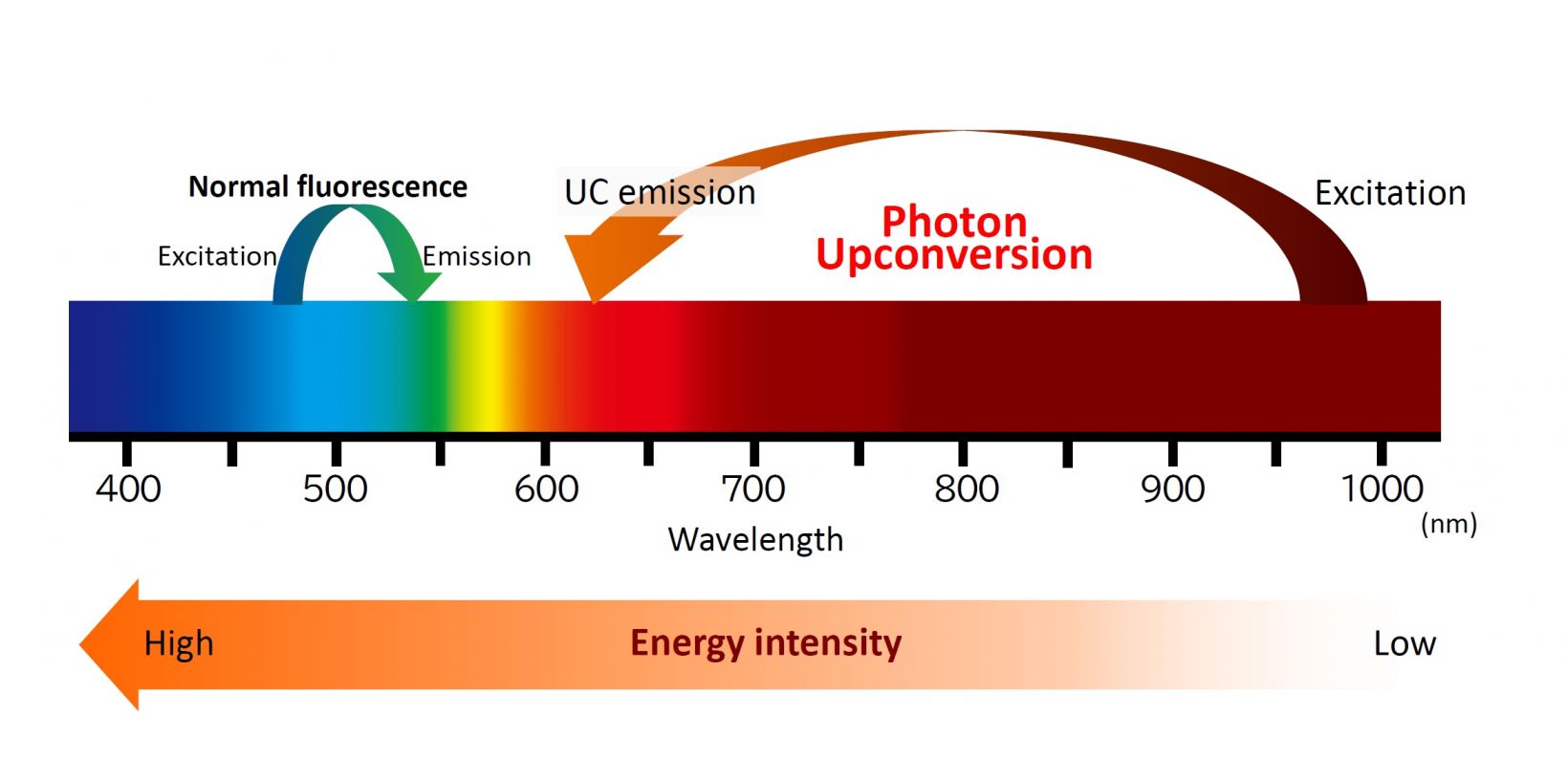
Figure 1. Conceptual diagram of photon upconversion
What Are the Benefits of Upconversion Imaging?
Fluorescence imaging with UCNPs uses an NIR wavelength laser (808 nm, 980 nm, etc.) for excitation, which enables imaging deeper into tissues and organisms with less scattering. Also, because longer wavelength light has lower energy, it causes less damage to the organism compared to excitation with higher energy visible light. UCNPs are also highly stable and do not photobleach easily, making them well suited for imaging both cells and living organisms.
Furthermore, the excitation for upconversion can be achieved with a CW diode laser instead of a high-power, pulsed laser. This enables researchers to perform experiments within the optimal NIR biological optical window* with the simplicity of a confocal microscope system, rather than a more complex and expensive multiphoton microscope system.
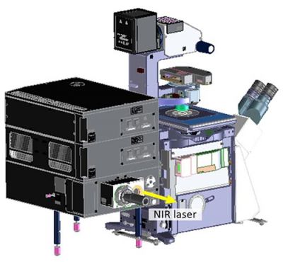
Figure 2. Example of the FLUOVIEW™ series confocal microscope with a laser introduction unit for upconversion
Examples of applications where upconversion can be used:
- Uncaging of caged compounds
- Activation of ion channels
- Nerve stimulation
- Luminescence resonance energy transfer (LRET)
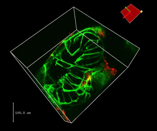
Figure 3. Zebrafish larvae, five days post-fertilization.
U87 glioma cells were coincubated with 5 ug/ml colominic acid coated UCNP for 12 hours, injected to 5 days post-fertilization zebrafish larvae and identified nanoparticle distribution in zebrafish.
Green channel: GFP expressed by the endothelial cells in transgenic zebrafish
Red channel (UCNP): 70 nm core-shell NaYbF4:Tm@NaYF4 nanoparticle coated by DSPE-PEG, no other labeling
Image courtesy of Dr. Yiqing Lu, School of Engineering, Macquarie University.
EVIDENT's Upconversion Solutions
Evident offers confocal laser-scanning microscope systems capable of upconversion imaging. We can propose a solution tailored to your experiment, including the type and number of lasers.
Please contact your local distributor for questions or to request a quote.
* Biological optical window: Near-infrared wavelength range (650 nm–1000 nm) where light easily penetrates living organisms.
References
▸Na Ren, Na Liang, Xin Yu, Aizhu Wang, Juan Xie, Chunhui Sun. Ligand-free upconversion nanoparticles for cell labeling and their effects on stem cell differentiation. Nanotechnology. 2020 Apr 3;31(14):145101.
▸Yanxiao Ao, Kanghua Zeng, Bin Yu, Yu Miao, Wesley Hung, Zhongzheng Yu, Yanhong Xue, Timothy Thatt Yang Tan, Tao Xu, Mei Zhen, Xiangliang Yang*, Yan Zhang, and Shangbang Gao. An Upconversion Nanoparticle Enables Near Infrared-Optogenetic Manipulation of the Caenorhabditis elegans Motor Circuit. ACS Nano 2019, 13, 3, 3373–3386.
▸Yong-Xiang Wu, Xiao-Bing Zhang, Dai-Liang Zhang, Cui-Cui Zhang, Jun-Bin Li, Yuan Wu, Zhi-Ling Song, Ru-Qin Yu, Weihong Tan. Quench-Shield Ratiometric Upconversion Luminescence Nanoplatform for Biosensing. Analytical Chemistry 2016, 88, 3, 1639–1646.
Related Content
Webinar: Introducing Upconversion Nanoparticles for Fluorescence Microscopy
Nanomaterials Brighten Up Life Science—Bioimaging Applications of Fluorescent Nanoprobes
Verifying the Tumor Labeling of a Novel Fluorescent Nanoprobe Using NIR Imaging