Verifying the Tumor Labeling of a Novel Fluorescent Nanoprobe Using NIR Imaging
Fluorescence imaging (FLI) is widely used for various biomedical imaging techniques in preclinical and clinical settings thanks to its advantages of high spatial and temporal resolution, high detection sensitivity, fast response time, and good operability. Applying near-infrared (NIR) capabilities to fluorescence imaging brings its own advantages of deep observation, low phototoxicity, and low interference with tissue autofluorescence. As a result, NIR fluorescence imaging has become a popular technique in medical research, especially in tumor treatment.
Fluorescence Imaging in the NIR-II Region
Compared with traditional fluorescent markers, such as fluorescent proteins and dyes, fluorescent nanoprobes can easily achieve NIR excitation imaging by adjusting their chemical composition and morphological size. Also, fluorescent nanoprobes can be further modified for localization, drug delivery, and multimodal detection.
To date, researchers have explored a variety of fluorescent probes to meet the growing demand for opportunities in precise disease diagnosis and treatment. Yet, most conventional fluorescent probes emit in the visible or NIR-I region (400–1000 nm). This greatly limits their applications in biomedical imaging due to drawbacks such as shallow tissue penetration depth and low spatial resolution.
As shown in recent studies, fluorescence imaging has been extended to the NIR-II region (1000–1700 nm). It displays great potential for deep tissue observation, such as high sensitivity and clarity, due to its imaging properties of low light scattering and low auto-fluorescence. As a result, this method can provide critical information in vivo that cannot be easily revealed using conventional fluorescence imaging.
Although fluorescence imaging technology continues to evolve, it is difficult to perfectly adapt single-modality optical imaging methods alone to the current requirements of in vivo, deep, ultra-sensitive, 3D, and high spatial and temporal resolution detection. As a result, there is an urgent need to develop multimodal optical detection systems.
Combining Bioimaging Methods to Improve Imaging Accuracy
Photoacoustic imaging (PAI) is a nondestructive biomedical imaging method that has been developed in recent years. When a pulsed laser is irradiated into biological tissues, the ultrasound signal generated by the biological tissues carries information about the tissues’ light absorption characteristics. As a result, dual-modality imaging combining NIR-II FLI and NIR-I PAI, which uses the advantages of each and compensates each other, can effectively improve imaging accuracy. It also has great application potential for efficient diagnosis.
To achieve high-quality imaging, the design of the probe is particularly important. Molecules with donor-acceptor-donor (D-A-D) structures have been attracting much attention for NIR-II molecular design due to their excellent biocompatibility, well-defined chemical structures, and customizable optical properties. To obtain NIR-II emission, the strongly electron-deficient unit, benzo[1,2-c:4,5-c']bis([1,2,5]thiadiazole (BBTD) was more widely used as an acceptor (Figure 1a).
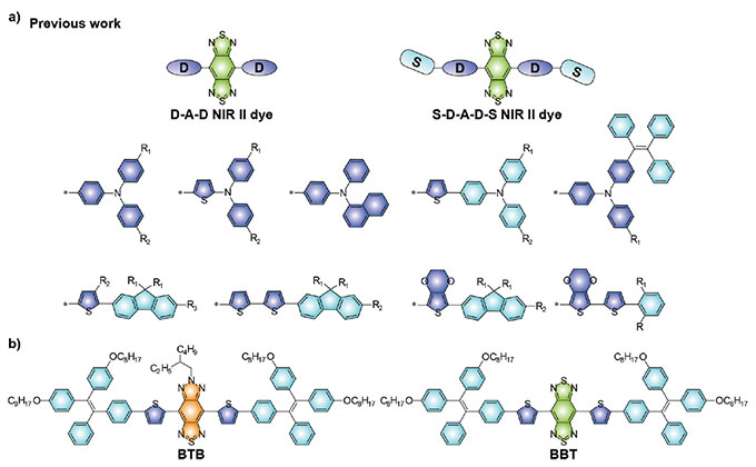
Figure 1. (a) Representative D-A-D type NIR-II fluorescent molecules based on BBTD receptors. (b) Chemical structures of the NIR-II fluorescent molecule BTB and the control fluorescent molecule BBT reported in the paper.
The quantum yields (QYs) of fluorescent of NIR-II molecules with a D-A-D structure are relatively low due to the presence of strong intramolecular charge transfer (ICT) effects. Many strategies have been developed to enhance QYs. These strategies include binding to dyes with hydrophobic structures of proteins, constructing fluorophores with aggregation-induced emission properties, introducing shielding units into molecular structures, and designing anti-burst molecules.
However, the QYs of most reported NIR-II fluorescent molecules with D-A-D structures are still relatively low due to their inherent ICT effects. In addition, BBTD requires relatively harsh synthesis conditions. This limits the development of more advanced NIR-II fluorescent molecules by functionalization. Therefore, exploring new receptor units is important to provide more alternative design strategies for the wide application of NIR-II fluorescent molecules.
Technical Requirements of NIR Fluorescent Probe Imaging
The in vitro/in vivo detection of NIR multimodal probes also poses new challenges for imaging instruments. Compared with conventional confocal imaging, NIR fluorescent probe imaging needs to meet these technical requirements:
- Most conventional confocal microscopes have excitation wavelengths between 400–650 nm. NIR imaging requires NIR lasers with >700 nm wavelengths.
- Conventional imaging optical components (e.g., scanning resonator, objective lens, and grating) are mostly only in the visible range for transmission/calibration. This cannot guarantee the efficiency and accuracy of NIR imaging.
- NIR detection requires a dedicated detector for >750 nm NIR. Ordinary detectors have very low detection efficiency in the NIR region.
Novel Fluorescent Nanoprobe for NIR-II Fluorescence and Photoacoustic Imaging
Together, Dr. Li Kai's group at SUSTech, Dr. Liu Jie's group at Nanjing University of Technology, and Dr. Chen Hao's group at Shanghai Institute of Pharmaceutical Sciences, Chinese Academy of Sciences, proposed a molecular design strategy of receptor engineering to obtain NIR-II fluorescent probes with high absorbance coefficients and quantum yields (Figure 1b). This strategy enabled functionalized probes for targeted detection of tumors in two- and three-dimensional mode conditions.
On November 10, 2021, the results of the study were published online in the Journal of Materials Chemistry B in a paper titled, "Receptor Engineering of Small Molecule Fluorophores for NIR-II Fluorescent and Photoacoustic Imaging."
We asked Dr. Yaxi Li, one of the first authors of the paper, to share the main findings and overall research experience:
1. What are the advantages of the novel fluorescent nanoprobe in bioimaging?
Conventional fluorescent probes do not target the tumor itself and are often retained at tumor sites by the enhanced permeability and retention effect (EPR effect), making them applicable for in vivo imaging of tumors. However, this passive enrichment approach requires a certain size of nanoparticles and is often inefficient. Therefore, to increase the enrichment of fluorescent probes at tumor sites and enhance the imaging effect for highly accurate diagnosis in vivo, we modified the nanoprobe surface with Arg-Gly-Asp (RGD) targeting peptide to obtain BTB-RGD NPs nanoprobes.
As illustrated in Figure 2a, the representative mouse injected with BTB-RGD NPs through the tall vein for 48 hours showed a significant increase in NIR-II fluorescence signal at the tumor site, and the signal reached the maximum value at 48 hours. In comparison, a control mouse injected with BTB NPs without targeting ability showed only a weak increase in fluorescence signal at the tumor site. Quantitative analysis (Figure 2b) showed the tumor site signals of a mouse injected with BTB-RGD NPs were always higher than those of the control group.
In addition to fluorescence imaging, we used photoacoustic tomography (PAT) to further confirm the overall targeting effect of BTB-RGD NPs on tumor sites (Figure 2c). The photoacoustic signal intensity at the tumor sites of the BTB-RGD NPs-injected mouse was also higher than that of the control-treated mouse—consistent with the NIR-II fluorescence imaging results.
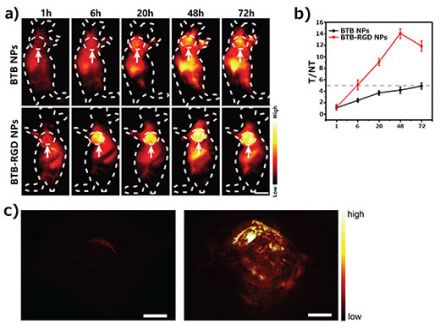
Figure 2. (a)Representative NIR-II fluorescence imaging images of 143B tumor-bearing mice taken 72 hours after injection of BTB-RGD NPs or BTB NPs under an 808 nm laser (140 mW cm-2) excitation; filter: 1000 nm long pass. Arrows indicate subcutaneous tumors. Number of mice per group (n) = 4. Scale bar = 5 mm. (b) T/NT ratio of 143B tumor imaged for 72 hours. Data are plotted as mean ± SD, n = 4. (c) Three-dimensional photoacoustic images of 143B tumors (730 nm excitation) in mice 48 hours after injection of BTB (left) or BTB-RGD NPs (right). Scale bar = 2 mm.
These imaging results successfully demonstrate the strong potential of our designed fluorescent probes for dual-mode bioimaging applications. On one hand, the high sensitivity of NIR-II fluorescence imaging was successfully used to achieve clear two-dimensional imaging of tumor margins. On the other hand, compared with fluorescence imaging, PAT has relatively lower temporal resolution and sensitivity but can provide superior spatial resolution and 3D visualization.
This dual-imaging approach will facilitate the reconstruction of volumetric images of tumor tissues, which is highly valued in clinical translational applications. For example, physicians can evaluate the 3D volume information of tumor tissue and perform fluoroscopic imaging for surgical tumor removal guided by a single dose of a contrast agent. Thus, integrating these two imaging modalities using a single contrast agent offers tremendous opportunities for precise vascular imaging and cancer treatment.
2. What technical difficulties did you encounter when imaging in the NIR region? How were they solved?
To demonstrate the targeting ability of BTB-RGD NPs, we first needed to determine their targeting ability on tumor cells through cellular assays. This required using confocal microscopy. However, most conventional confocal microscopes are equipped with lasers limited to the visible region. Therefore, to validate the NIR-II targeting probe, the group could only indirectly demonstrate the targeting ability by replacing the probe with a similar structure in the visible region. However, this approach cannot visually demonstrate the targeting ability of the NIR-II probe itself. It also increases the workload of probe preparation during the experiment.
Therefore, a confocal microscope equipped with an NIR laser is particularly important. To solve this difficulty, we used a FLUOVIEW™ FV3000 confocal laser scanning microscope upgraded with the FV3000 Red near-infrared (NIR) imaging solution. Using the FV3000 Red system, we successfully determined the targeting ability of BTB-RGD NPs on tumor cells. Figure 3 shows that after incubation with 40 μg mL-1 BTB- RGD NPs alone, the fluorescence signal of cancer cells was significantly higher than that of the control group of BTB NPs. In contrast, the fluorescence intensity of normal 3T3 cells incubated with BTB-RGD NPs did not differ significantly from that of BTB NPs treatment. These results suggest that nanoparticles after functionalization with RGD can promote their specific targeting to tumor cells overexpressing αvβ3 integrin.
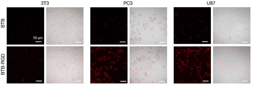
Figure 3. Confocal images of 3T3, PC3, and U87 cells after incubation with 40 μg mL-1 of BTB-RGD or BTB NPs for 4 hours. Excitation wavelength: 730 nm, filter: 760–890 nm, scale bar = 50 μm.
In previous experiments, tumor labeling could only be confirmed indirectly. This was done by replacing the probe with a similar structure in the visible range, as the system’s observation range was limited to visible light only. By using the FV3000 Red system in this experiment, we could successfully measure the labeling of BTB-RGD NPs on tumor cells without replacing the probe.
Near-Infrared (NIR) Solutions for Confocal Microscopy
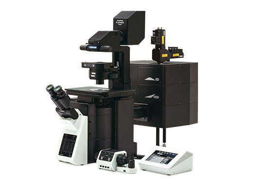
The FV3000 Red system extends the FV3000 microscope’s wavelength detection capabilities to the NIR region. By upgrading each imaging module for NIR detection, including the laser, optics, objective lens, and detector, the FV3000 Red system provides a specialized solution for more sensitive and accurate multi-color NIR imaging.
| FV3000 Red features:
|
| AcknowledgementsThis application note was prepared with the help of the following researcher: Dr. Kai Li, Southern University of Science and Technology |

References: 1.V. J. Yao, S. D’Angelo, K. S. Butler, C. Theron, T. L. Smith, S. Marchio, J. G. Gelovani, R. L. Sidman, A. S. Dobraff, C. J. Brinker, A. R. M. Bradbuty, W. Arap. and R. Pasqualini, Ligandtargeted theranostic nanomedicines against cancer, J. Control. Release 2016, 240, 267-286. 2. B. Guo, Z. Sheng, D. Hu, C. Liu, H. Zheng, B. Liu, Through Scalp and Skull NIR-II Photothermal Therapy of Deep Orthotopic Brain Tumors with Precise Photoacoustic Imaging Guidance, Adv. Mater. 2018, 30, 1802591. 3. G. L. Bagnato, N. Irrera, G. Pizzino, D. Santoro, W. N. Roberts, G. Bagnato, G. Pallio, M. Vaccaro, F. Squadrito, A. Saitta, D. Altavilla, and A. Bitto, Dual αvβ3 and αvβ5 blockade attenuates fibrotic and vascular alterations in a murine model of systemic sclerosis, Clin. Sci. 2018, 132, 231-242. 4. N. Zoppi, N. Chiarelli, V. Cinquina, M. Ritelli, and M. Colombi, GLUT10 deficiency leads to oxidative stress and non-canonical αvβ3 integrin-mediated TGFβ signalling associated with extracellular matrix disarray in arterial tortuosity syndrome skin fibroblasts, Hum. Mol. Genet. 2015, 24, 6769-6787. 5. Y. Li, Z. Li, D. Hu, S. Wang, M. Zha, S.-B. Lu, Z. Sheng, and K. Li, Targeted NIR-II emissive nanoprobes for tumor detection in mice and rabbits, Chem. Commun. 2021, 57, 6420-6423.
Products related to this application
Sorry, this page is not
available in your country.