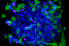
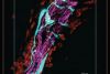
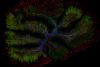
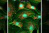
Fluorescence imaging visualizes intracellular structures, particularly protein and molecular structures, using fluorescent proteins or dyes. To acquire high-quality fluorescence images, specialized devices and optical equipment are required, and they need to be properly set up.
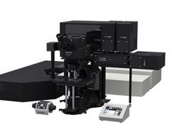
Our fluorescence imaging systems use UIS2 optics, a series of high-performance objectives that offer both a high numerical aperture (NA) and precise compensation of spherical and chromatic aberration. Our fully motorized fluorescence imaging systems provide easy, one-click system setup and multidimensional image acquisition, such as multicolor, multiple image alignment (MIA), and Z-stack, with a simple workflow.
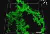
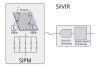
We also offer fluorescence imaging software that enables high-resolution fluorescence image acquisition through powerful image processing techniques, such as deconvolution. The software gives you access to a wide range of analysis methods, including intensity profiling, automatic object measurement, and classification.
Browse fluorescence imaging applications, as well as our range of systems, cameras, and software to build your optimal fluorescence imaging solution.
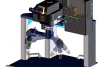
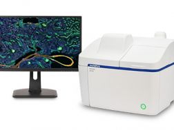
The APEXVIEW™ APX100 benchtop fluorescence microscope makes it fast and simple to acquire expert-quality microscope images. Built with our renowned optics, an intuitive user interface, a powerful AI, and a suite of smart features, the APX100 system combines ease of use with high-quality image data to fit your research needs.
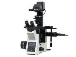
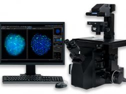
Optimized for basic multicolor fluorescence imaging and routine experiments, the IXplore Standard system is easy to operate and ergonomically designed. Even with standard cell culture vessels, it captures high-quality, publication-worthy images while providing accurate and repeatable results at high magnifications. The IXplore Standard system’s simplified workflow and ease of use facilitate a wide range of standard imaging applications.
High repeatability and accuracy for standard imaging tasks
Benefit from the same optical capabilities found in high-end IXplore systems
Easily upgrade to encoded functionality to boost the reproducibility of experiments
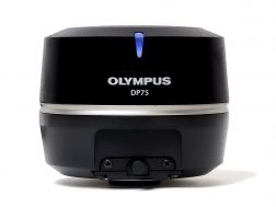
The high-performance DP75 digital microscope camera makes it easy to capture high-resolution brightfield or fluorescence images using a single color camera. It simplifies your microscopy imaging, so you can focus more on your work.
Integrated TruAI denoising maximizes the camera’s image quality in real time
Exceptional color reproduction, making your images as vivid as looking through the microscope oculars
Supports multiple staining combinations and wavelengths up to 1000 nm with a switchable infrared (IR) cut filter
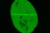
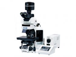
The monochrome DP23M microscope digital camera is a cost-effective solution for high-quality fluorescence imaging. Even for dim samples, its high sensitivity and resolution deliver bright images, easing protein expression checks, observation using near-infrared (NIR) dyes, and other routine fluorescence imaging tasks.
Providing intuitive operations and a seamless workflow, cellSens software’s user interface is customizable so you control the layout. Offered in a range of packages, cellSens software provides a variety of features optimized for your specific imaging needs. Its Graphic Experiment Manager and Well Navigator features facilitate 5D image acquisition. Achieve improved resolution through TruSight™ deconvolution and share your images using Conference Mode.
Improve experiment efficiency with TruAI™ deep-learning segmentation analysis, providing label-free nuclei detection and cell counting
Modular imaging software platform
Intuitive application-driven user interface
Broad feature set, ranging from simple snapshot to advanced multidimensional real-time experiments