Evident LogoOlympus Logo

Drug Discovery

Life Science Research Application

Applications: Drug Discovery

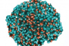
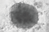
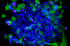
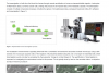
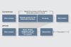
Pharmaceutical companies use cell models for their research as an alternative to animal experiments and for more precise evaluation of drug efficacy. In recent years, 3D cell models called spheroids, organoids, and “body-on-a-chip” have been engineered and applied to pharmaceutical research. We provide not only conventional brightfield observation solutions but also confocal microscopes and multiphoton excitation microscopes for deep specimen image acquisition, enabling high-resolution observation of 3D structures. To prepare for cell model construction, it is important to keep the quality and number of cells constant, so we also offer reliable cell culture solutions. Our slide loader system and analysis solutions can acquire high-speed, high-resolution images of an entire section of tissue for high-precision pharmacology and drug efficacy analysis.
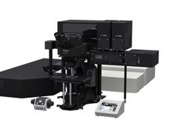
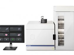
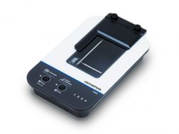
Solutions for Drug Discovery - Build Your Configuration

Successfully added visible items to your Bookmarks list

View Bookmarks

Microscope System/Frame

FV4000Show Description
FV4000

  • Game-changing dynamic range for imaging from the macro scale to subcellular structures
  • Multiplex up to six channels simultaneously with TruSpectral technology
  • Redesigned high-speed, high-resolution scanners for fixed and live cell imaging
  • Improved depth and photosensitivity with pioneering NIR capabilities and renowned optics
  • Peace of mind with the reliable, repeatable SilVIR detector
  • Industry leading* ten laser lines with a broader spectral range from 405 nm to 785 nm

*As of October 2023.

FV4000

Confocal Laser Scanning Microscope
ChooseSelected  
FV4000MPEShow Description
FV4000MPE

  • Acquire accurate, quantitative image data from the macro scale to subcellular structures
  • Obtain more information from a single multicolor image
  • Monitor neuron and other essential dynamics with high-speed imaging

FV4000MPE

Multiphoton Laser Scanning Microscope
ChooseSelected  
APX100Show Description
APX100

The APEXVIEW APX100 all-in-one microscope makes it fast and simple to acquire expert-quality microscope images. Built with renowned Olympus optics, an intuitive user interface, a powerful AI, and a suite of smart features, the APX100 system combines the ease of use of an all-in-one-microscope with high-quality image data to fit your research needs.

  • Stay focused on your research
  • Publication-quality images in a few clicks
  • Fast, efficient data management

APX100

Digital Imaging System
ChooseSelected  
VS200Show Description
VS200
  • Outstanding image quality for whole slide imaging, from 2X to 100X, including oil scanning
  • Accommodates multiple slide sizes and observation methods
  • SILA optical sectioning device enables you to obtain high-contrast images free from blur across the entire slide
  • Simplified and powerful workflow, from brightfield to fluorescence multiplexing
  • Uniquely configurable software and hardware, from manual brightfield scanning to AI recognition and fully automated scanning

VS200

Research Slide Scanner
ChooseSelected  
IXplore ProShow Description
IXplore Pro

The IXplore Pro automated system acquires panoramic, multichannel images without a complex workflow. Easing experiment setup, the Graphical Experimental Manager offers fully automated multidimensional observation capabilities. The microscope’s high-precision ultrasonic stage, automated focusing, and bright, uniform fluorescence illumination facilitate image stitching applications.  

  • Automated multidimensional observation (X, Y, Z, T, wavelength, and positions)
  • Boost your statistics with multiwell plate screening
  • Acquire fluorescence panoramic images of large samples, such as brain slices
  • Create 3D optical sections and enhance resolution with TruSight™ deconvolution

IXplore Pro

Automated Microscope System
ChooseSelected  
IXplore LiveShow Description
IXplore Live

Designed to reduce photobleaching and phototoxicity, the IXplore Live system is optimized for physiological experiments involving live cell and tissue observation. Offering precise environmental control and enhanced rigidity, it supports long-term cell viability and stability for time-lapse imaging applications, such as in cancer, stem cell, and brain research.

  • Maintain focus accurately and reliably in time-lapse experiments with TruFocus™ Z-drift compensation system
  • Discover the real morphology of your cells with Olympus silicone immersion optics
  • Real-time controller helps limit cell disturbance, enabling physiologically relevant data

IXplore Live

Live Cell Imaging Microscope System
ChooseSelected  
IXplore SpinShow Description
IXplore Spin

The IXplore Spin system features a spinning disk confocal unit that enables fast 3D image acquisition, a large field of view, and prolonged cell viability in time-lapse experiments. Researchers can use it to perform rapid 3D confocal imaging with high resolution and contrast at greater depths for imaging into thicker samples. The spinning disk also helps to cut down on photobleaching and phototoxicity of samples upon excitation.

  • Real-time controller (U-RTCE) helps optimize the device’s speed and precision during automated acquisition
  • TruFocus™ Z-drift compensation system maintains focus for each frame
  • Precise 3D imaging with improved light collection using X Line™ objectives
  • Upgrade to the IXplore SpinSR super resolution system as your research progresses

IXplore Spin

Confocal Imaging Microscope System
ChooseSelected  
scanRShow Description
scanR

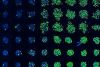
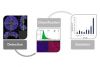
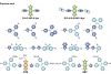
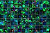
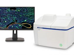
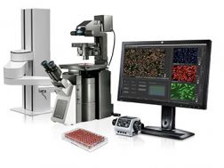
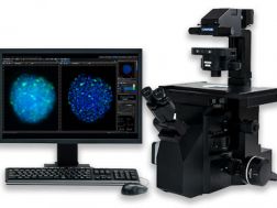
Achieve fully automated image acquisition and data analysis of biological samples using the scanR high-content screening station. Design individualized assays for cell cycle, protein localiazation, intracellular transport and more.  Modular hardware is compatible with a range of additional systems, including spinning disk confocal, robot loading, incubation, TIRF, and FRAP systems.

  • Fast and precise image acquisition and analysis
  • Image cytometry based approach enables easy and detailed visualization of results
  • Expand your capabilities with modules such as self-learning AI, kinetic parameter measuring, high-speed 3D deconvolution, and more

scanR

High-Content Screening Station for Life Science
ChooseSelected  
CKX53Show Description
CKX53

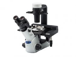
The CKX53 microscope eases the cell and tissue culture workflow, simplifying steps such as live cell observation, cell sampling and handling, image capture, and fluorescence observation. Its integrated phase contrast system, compact, ergonomic design, and stable performance enable simple, efficient cell observation. The universal sample holder and expandable stage accommodate a wide variety of cell culture container types and sizes.

  • Precentered phase contrast
  • Inversion contrast (IVC) technique provides clear three-dimensional views
  • Fluorescence with a 3-position slider
  • View multilayer tissue flasks up to 190 mm (7.5 in.) in height up thanks to the detachable condenser

CKX53

Compact Cell Culture Microscope
ChooseSelected  

Cameras

DP75Show Description
DP75

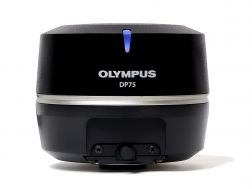
The high-performance DP75 digital microscope camera makes it easy to capture high-resolution brightfield or fluorescence images using a single color camera. It simplifies your microscopy imaging, so you can focus more on your work.

  • Integrated TruAI denoising maximizes the camera’s image quality in real time
  • Exceptional color reproduction, making your images as vivid as looking through the microscope oculars
  • Supports multiple staining combinations and wavelengths up to 1000 nm with a switchable infrared (IR) cut filter

DP75

Microscope Digital Camera
ChooseSelected  
XM10Show Description
XM10
  • 1.4-megapixel monochrome cooled CCD
  • Low noise and high sensitivity
  • Fast Live image speed

XM10

Digital Microscope Camera
ChooseSelected  
DP23Show Description
DP23

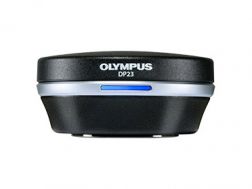
Enabling fast, easy capture of high-quality images that can be clearly observed on a large screen, the DP23 microscope digital camera eases routine life science and clinical research, conferencing, or teaching. Integrate it seamlessly into your microscopy workflow and easily share or stream images.  

  • Share images using the DP23-AOU network solution
  • Clearly observe live images on a large screen
  • Fast, high-quality imaging for conferences and teaching

DP23

Microscope Digital Camera
ChooseSelected  
DP28Show Description
DP28

Providing color accuracy and 4K resolution, the DP28 digital microscope camera’s powerful features and wide field of view capture images that enhance tasks such as conferencing, teaching, and clinical research. Integrate it seamlessly into your microscopy workflow for improved work efficiency and image quality. 

  • 4K resolution
  • Color reproduction that rivals the human eye
  • 8.9-megapixel CMOS sensor

DP28

Microscope Digital Camera
ChooseSelected  
Not ApplicableShow Description
Not Applicable

Software

cellSensShow Description
cellSens

Providing intuitive operations and a seamless workflow, cellSens software’s user interface is customizable so you control the layout. Offered in a range of packages, cellSens software provides a variety of features optimized for your specific imaging needs. Its Graphic Experiment Manager and Well Navigator features facilitate 5D image acquisition. Achieve improved resolution through TruSight™ deconvolution and share your images using Conference Mode.

  • Improve experiment efficiency with TruAI™ deep-learning segmentation analysis, providing label-free nuclei detection and cell counting
  • Modular imaging software platform
  • Intuitive application-driven user interface
  • Broad feature set, ranging from simple snapshot to advanced multidimensional real-time experiments

cellSens

Imaging Software
ChooseSelected  
NoviSightShow Description
NoviSight

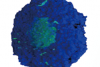
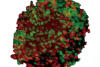
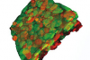
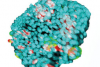
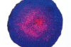
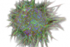
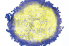
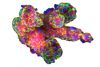
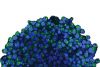
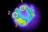
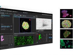
NoviSight 3D cell analysis software provides statistical data for spheroids and 3D objects in microplate-based experiments. Use it to quantify cell activity in 3D, easily capture rare cell events, obtain accurate cell counts, and improve detection sensitivity. NoviSight software works with a range of imaging techniques, including point-scan confocal imaging, two-photon imaging, spinning disk confocal imaging, and super resolution live cell imaging.

  • Fast 3D image recognition from whole structures to subcellular features
  • Accurate statistical analysis
  • Equipped with a variety of ready-to-use default assays or easily design your own

NoviSight

3D Cell Analysis Software
ChooseSelected  
Not ApplicableShow Description
Not Applicable

Cell Culture Equipment

CM30Show Description
CM30

Remotely monitor, analyze, and share your cell cultures’ health, cell count, and confluency using the reliable quantitative data provided by the automated CM30 incubation monitoring system. The system enables label-free observation, reduces the risk of damage to your cultures, and standardizes your culture workflow.

  • Automatically collects quantitative data on the health and confluency of your cultures
  • Monitor, analyze, and share your cultures' progress remotely from a PC or tablet
  • Equipped with oblique epi-illumination for label-free observation

CM30

Incubation Monitoring System
ChooseSelected  
Not ApplicableShow Description
Not Applicable

Sorry, this page is not
available in your country.

Sorry, this page is not available in your country