Not Available in Your Country
Sorry, this page is not
available in your country.
Overview
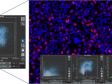
 | Modular High-Content Screening Station for Life SciencesThe scanR modular microscope-based imaging platform provides fully automated image acquisition and data analysis of biological samples through deep-learning technology. |
|---|
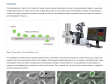
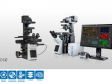
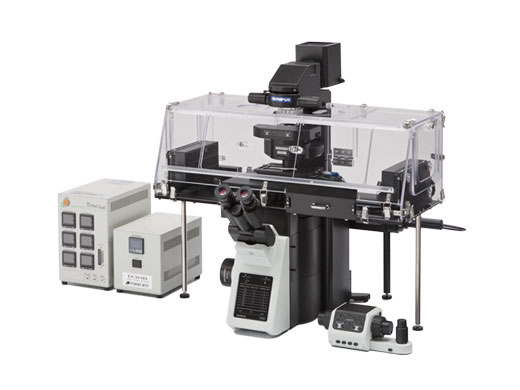
Flexible, Modular HardwareThe scanR screening station combines the modularity and flexibility of a microscope-based setup with the automation, speed, and throughput demanded by high-content screening. Well-suited for standard assays and assay development, the modular design makes the scanR station adaptable to R&D lab applications or multiuser environments. |
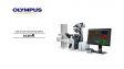
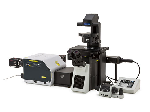
Spinning Disk Confocal System
|
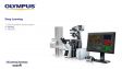
Robot Loading System
|
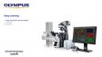
Incubation System
|
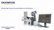
TIRF and FRAP System (with cellSens Software)
|
Related Videos | Comprehensive SoftwareThe system’s flexible design enables it to meet the requirements for quantitative imaging and image analysis in modern cell biology, molecular biology, systems biology, and medical research.
|
|---|
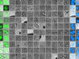
Meets the Needs of Many AssaysThe scanR system excels in drug discovery applications, including showing the biochemical effects of compounds on the cellular level and drug-induced changes at gene expression levels. The solution can measure apoptosis, micronuclei, or DNA fragmentation (comet assays) and covers a wide range of screening applications:
|
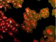
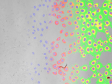
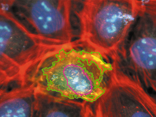
Genome-wide screen on cell arrays to identify novel genes involved in intracellular transport machinery. Image courtesy of Dr R. Peperkok, EMBL, Heidelberg, Germany. |
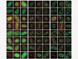
Chlamydia trachomatis infection assay. Image courtesy of Dr S. Hess, Max Planck Institute (MPI) for Infection Biology, Berlin, Germany. |
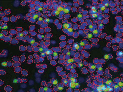
Segmentation of binucleated cells and micronuclei counting. Image courtesy of Department for In-Vitro Toxicology, Fraunhofer Institute Toxicology and Experimental Medicine (ITEM), Hanover, Germany. |
Flexible Module OptionsThe scanR solution not only satisfies the specific speed, endurance, and reliability requirements of a fully automated high-content screening system but also provides unmatched flexibility and adaptability with extensive expansion capabilities. This enables the scanR system to meet the specifications of a wider range of applications and budgets. Add modules with the following capabilities to your system:
Contact application specialists to customize your system for your applications
|
Need assistance? |
Applied Technologies
Gating and Classification
|
A hierarchical gating approach enables intuitive selection of populations, which may also be visualized in galleries. |
|---|
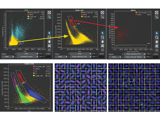
Get Started QuicklyThe included pre-trained neural network models enable you to start using the AI fast. Using the pre-trained models, you can begin detecting nuclei and cells in most standard conditions. Even confluent cells and dense nuclei can be reliably distinguished. Control and validation measures are built in to help ensure the accuracy and robustness of the AI analysis results. |
|
|
|
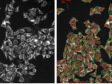
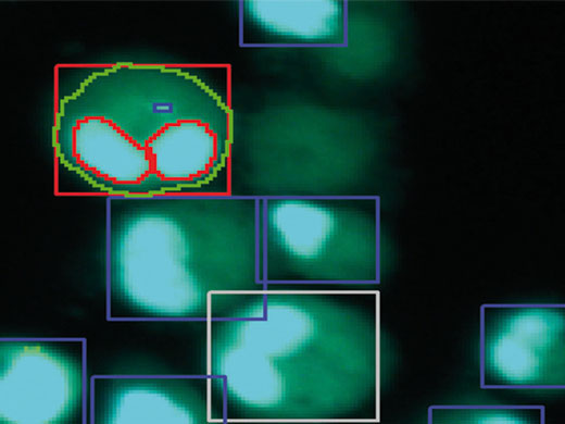
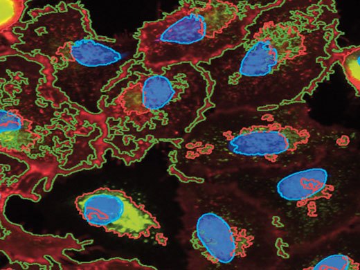
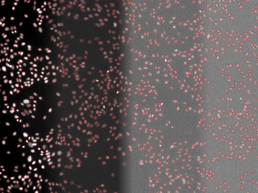
Accurate object segmentation: raw data (left), standard threshold segmentation (middle), TruAI instance segmentation (right). Instance segmentation reliably separates difficult-to-distinguish objects that are very close together, such as cells or nuclei in colonies or tissue. |
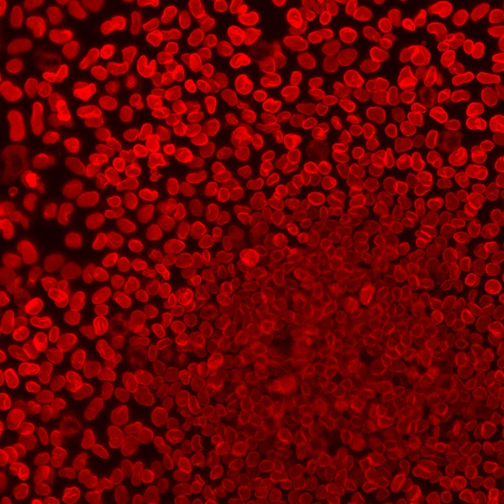
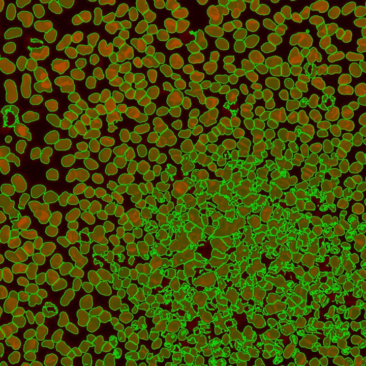
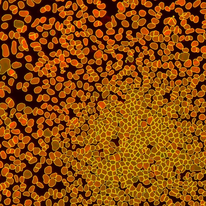
Image screenshot detail following data acquisition by scanR demonstrating the detection and separation of labels. Courtesy of Dr. R. Pepperkok, EMBL Heidelberg, Germany. | Object Detection and Analysis
|
|---|
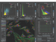
Immediate Quality ControlImages and objects are reciprocally linked to their related data points:
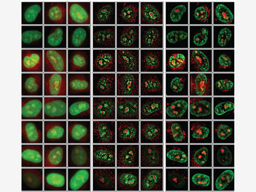
Create a gallery view of all images of a selected or gated data population to enable direct, visual comparison of larger image sets with relevant information. |
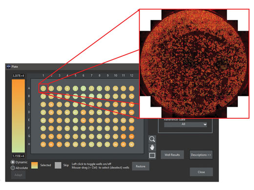
Results are visualized in heatmaps or exported to tables. Easily display an overview of full wells. |
|---|
Related Videos | Multilevel AcquisitionAfter an initial prescan, the scanR analysis software can identify all the potential objects of interest. In an automated workflow, the analysis results are used to selectively scan the objects of interest in a second, targeted screen. |
|---|
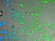
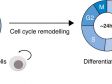
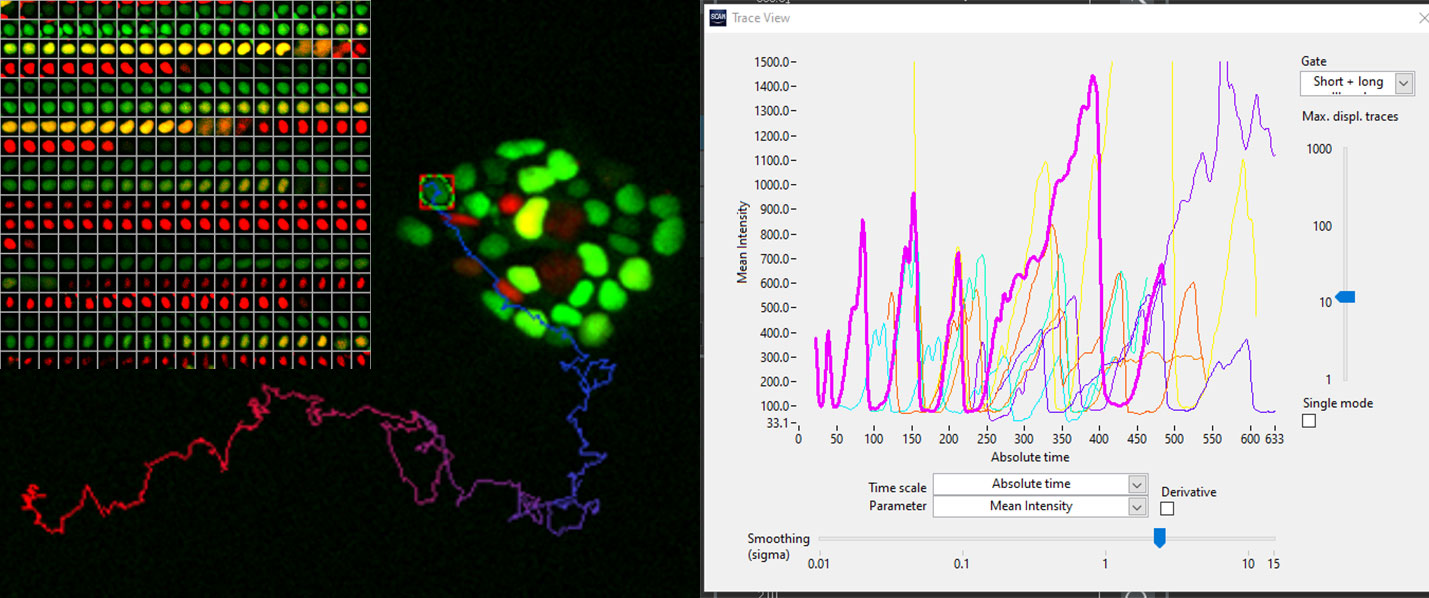
Measuring Kinetic Parameters with the Kinetic Module
|
hES cells expressing FUCC (CA) biosensor. Courtesy of Dr. Silvia Santos, The Francis Crick Institute, London, UK. |
|---|
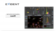
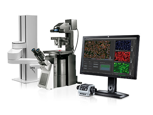
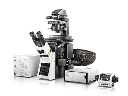
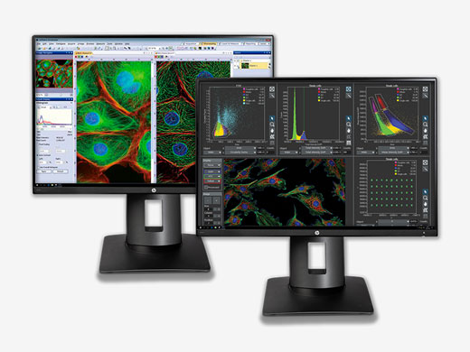
| Combines High-End Imaging and High-Content Analysis
|
|---|
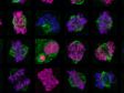
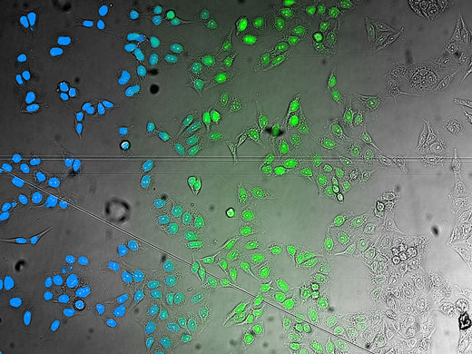
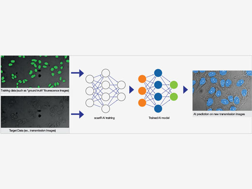
Self-Learning MicroscopySelf-learning microscopy opens new horizons in high-content analysis. Applications range from previously impossible image segmentation and classification tasks to quantitative analysis of extremely low signal levels, simplifying staining protocols, label-free analysis, and more. |
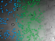
Example workflow using self-learning microscopy to generate an AI model for label-free analysis of challenging brightfield images. The cell nuclei of Hela cells are GFP-labeled for the training phase to show the system how to analyze the brightfield images. |
Example application: Robust segmentation of cell nuclei at different signal levels, enabling a dramatic reduction of light exposure for quantitative analysis. |
Related VideosThe user has full control of the training experiment design. | Related VideosMany challenging analysis conditions can be covered during the training phase. | Related VideosThe learned AI analysis protocol can be validated in depth and with ease with the software’s unique data exploration and analysis interface. |
Need assistance? |
Specifications
| Microscope Frame |
Olympus IX83 inverted microscope, one or two decks
Motorized stage, Märzhäuser SCAN IM 120 × 80 for the Olympus IX83 microscope |
|---|---|
| Fluorescence LED Options |
Lumencor SpectraX: 6 independent LED channels
CoolLED pE-300ultra: 3 independent LED channels Application-optimized bandpass filters Lifetime > 20,000 hours |
| Transmitted-Light Illumination Options |
Transmitted-light illumination for visual inspection only (no transmitted-light screening)
Transmitted-light illumination for screening and visual inspection including fast shutter (transmitted-light screening supported) Optional DIC (differential interference contrast) or phase contrast |
| Hardware Control and System Synchronization (optional) |
Real-time controller with additional CPU, independent of the OS of the imaging PC
Temporal resolution: 1 ms Timing precision: < 0.01 ms Hardware-synchronized multitask acquisition (illumination control, emission filters, shutters, etc.) Precise camera control via external trigger |
| Camera Options |
Hamamatsu ORCA-Flash 4.0 V3, high-sensitivity cooled sCMOS camera with large 18.8 mm (0.74 in.) sensor chip
Hamamatsu ORCA-Flash 4.0 LT, an economic sCMOS camera with large 18.8 mm (0.74 in.) sensor chip Hamamatsu ORCA-Fusion, sCMOS camera with large 21.2 mm (0.83 in) sensor chip Hamamatsu ORCA-Fusion BT, ultra-low noise sCMOS camera with large 21.2 mm (0.83 in.) sensor chip |
|
Objective Options
(supports Olympus X Line objectives) |
Objectives for “thin” (0.1 mm–0.2 mm [0.004 in.–0.008 in.]) substrates, cover slips, and glass bottom plates (2X, 4X, 10X, 20X, 40X, 60X, 100X)
Objectives for “thick” (~1 mm [0.04 in.]) substrates, plastic bottom plates, and slides (2X, 4X, 10X, 20X, 40X, 60X, 100X) Phase contrast objectives for “thin” (0.1 mm–0.2 mm [0.004 in.–0.008 in.]) substrates, coverslips, and glass bottom plates (10X, 20X, 40X) Phase contrast objectives for “thick” (~1 mm [0.04 in.]) substrates, coverslips, and glass bottom plates (10X, 20X, 40X) |
| Filter Sets |
Single-band filter sets (specifications as requested)
Multiband filter sets (specifications as requested) |
|
scanR System
Software |
2 independent software modules: scanR acquisition software and scanR analysis software
Shading correction workflow to compensate for shading and optimize spatial intensity homogeneity during and post-acquisition The software modules can be installed on the same or on different workstations (Windows 10 64-bit) |
|
scanR Acquisition
Software |
Workflow-oriented configuration and user interface
Powerful software autofocus procedures that can be combined with an optional IR laser hardware autofocus function, 2-step coarse and fine autofocus, object-based autofocus, or image-based autofocus Plate manager with predefined formats (slides, multiwell plates) and editing interface to create and edit customized formats (spotted arrays) Shading correction to compensate for shading and optimize spatial intensity homogeneity Time-lapse screening, Z-stack screening, multicolor screening (unlimited number of acquisition channels) Support for integration into automated sample-preparation lines, ex., scriptable interfaces for liquid handling |
|
scanR Analysis
Software |
Automated image and data analysis for standard assays and assay development
Online and offline multicore analysis Image processing, image analysis, particle detection, and parameter extraction and calculation Cytometric data exploration, analysis, gating, and classification Powerful and flexible gating concept including automated population detection Direct link between data points, objects, and images Assays-based work flow and advanced scientific assay development functionality |
| Computer | Imaging computer (latest generation PC) , Windows 10 64 bit |
| Additional Options |
scanR AI deep-learning solution
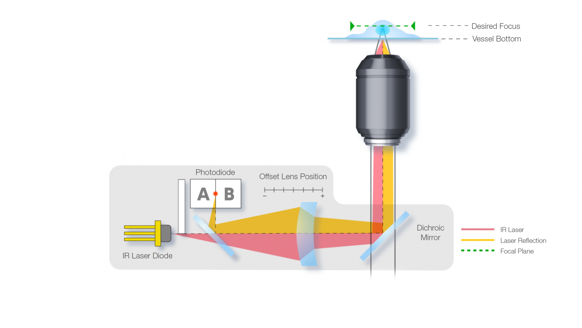
Time-lapse kinetic analysis module—a unique cytometric approach to better analyze and understand live-cell dynamics 3D deconvolution module (64-bit operating system required) Confocal option with Yokogawa CSU-W1 with one or two cameras Two-camera simultaneous acquisition IR laser hardware autofocus function based on the ZDC of the Olympus IX83 microscope series Plate-loading robot Encoded magnification changer IX3-CAS Fast-emission filter wheel for high-speed imaging in “Sedat” configuration Additional scanR analysis workstation Second license for scanR analysis software scanR Analysis Viewer |
| Two Systems in One Setup | Can be combined with cellSens live cell imaging software for full versatility of a high-end imaging system |