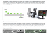
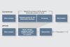
An effective cell culture process lays the foundation for success in many application areas throughout the life science and the pharmaceutical industry, such as cancer research, genome editing, stem cell research, and regenerative medicine. With improved image quality and easy handling, our cell culture microscope system delivers stable performance and a more efficient cell culture process for a variety of cell culture needs including live cell observation, cell sampling and handling, image
capture, and fluorescence observation. To accelerate the cell culture process, a cell counter offers easy and smooth operation when used in conjunction with a microscope for quick live imaging and accurate cell count of cultured cells. An efficient flow of cell observation and counting can be accomplished with our cell culture solutions.
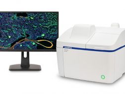
The APEXVIEW™ APX100 benchtop fluorescence microscope makes it fast and simple to acquire expert-quality microscope images. Built with our renowned optics, an intuitive user interface, a powerful AI, and a suite of smart features, the APX100 system combines ease of use with high-quality image data to fit your research needs.
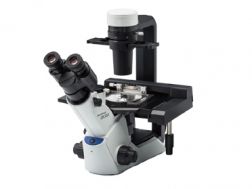
The CKX53 microscope eases the cell and tissue culture workflow, simplifying steps such as live cell observation, cell sampling and handling, image capture, and fluorescence observation. Its integrated phase contrast system, compact, ergonomic design, and stable performance enable simple, efficient cell observation. The universal sample holder and expandable stage accommodate a wide variety of cell culture container types and sizes.
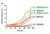
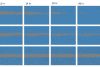
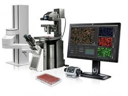
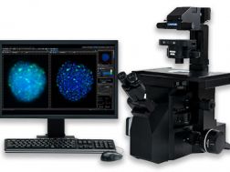
Achieve fully automated image acquisition and data analysis of biological samples using the scanR high-content screening station. Design individualized assays for cell cycle, protein localiazation, intracellular transport and more. Modular hardware is compatible with a range of additional systems, including spinning disk confocal, robot loading, incubation, TIRF, and FRAP systems.
Fast and precise image acquisition and analysis
Image cytometry based approach enables easy and detailed visualization of results
Expand your capabilities with modules such as self-learning AI, kinetic parameter measuring, high-speed 3D deconvolution, and more
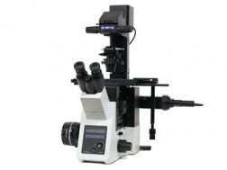
Optimized for basic multicolor fluorescence imaging and routine experiments, the IXplore Standard system is easy to operate and ergonomically designed. Even with standard cell culture vessels, it captures high-quality, publication-worthy images while providing accurate and repeatable results at high magnifications. The IXplore Standard system’s simplified workflow and ease of use facilitate a wide range of standard imaging applications.
High repeatability and accuracy for standard imaging tasks
Benefit from the same optical capabilities found in high-end IXplore systems
Easily upgrade to encoded functionality to boost the reproducibility of experiments
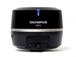
The high-performance DP75 digital microscope camera makes it easy to capture high-resolution brightfield or fluorescence images using a single color camera. It simplifies your microscopy imaging, so you can focus more on your work.
Integrated TruAI denoising maximizes the camera’s image quality in real time
Exceptional color reproduction, making your images as vivid as looking through the microscope oculars
Supports multiple staining combinations and wavelengths up to 1000 nm with a switchable infrared (IR) cut filter
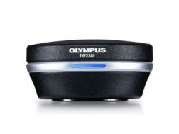
The monochrome DP23M microscope digital camera is a cost-effective solution for high-quality fluorescence imaging. Even for dim samples, its high sensitivity and resolution deliver bright images, easing protein expression checks, observation using near-infrared (NIR) dyes, and other routine fluorescence imaging tasks.
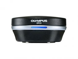
Enabling fast, easy capture of high-quality images that can be clearly observed on a large screen, the DP23 microscope digital camera eases routine life science and clinical research, conferencing, or teaching. Integrate it seamlessly into your microscopy workflow and easily share or stream images.
Share images using the DP23-AOU network solution
Clearly observe live images on a large screen
Fast, high-quality imaging for conferences and teaching
Providing intuitive operations and a seamless workflow, cellSens software’s user interface is customizable so you control the layout. Offered in a range of packages, cellSens software provides a variety of features optimized for your specific imaging needs. Its Graphic Experiment Manager and Well Navigator features facilitate 5D image acquisition. Achieve improved resolution through TruSight™ deconvolution and share your images using Conference Mode.
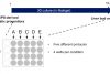
Improve experiment efficiency with TruAI™ deep-learning segmentation analysis, providing label-free nuclei detection and cell counting
Modular imaging software platform
Intuitive application-driven user interface
Broad feature set, ranging from simple snapshot to advanced multidimensional real-time experiments
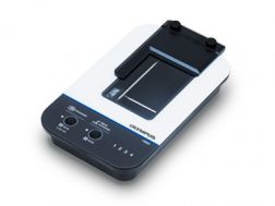
Remotely monitor, analyze, and share your cell cultures’ health, cell count, and confluency using the reliable quantitative data provided by the automated CM30 incubation monitoring system. The system enables label-free observation, reduces the risk of damage to your cultures, and standardizes your culture workflow.
Automatically collects quantitative data on the health and confluency of your cultures
Monitor, analyze, and share your cultures' progress remotely from a PC or tablet
Equipped with oblique epi-illumination for label-free observation