Not Available in Your Country
Sorry, this page is not
available in your country.
Overview
 | Automated Light Sheet System for Imaging FacilitiesThe Alpha3 Facility Edition by PhaseView is a sophisticated light sheet microscope designed for core facilities. It combines optimal image quality, an intuitive user experience, and the flexibility for scientific imaging instruments. As a fully automated system, the Alpha3 Facility Edition automatically configures the microscope when changing the magnification so you can focus on imaging. The Alpha3 Facility Edition comes with the intuitive QTSPIM software for image acquisition at maximum speed and the LINDA 3D viewer to ease sample exploration. |
|---|
Key Benefits of the Alpha3 Facility Edition
| Key Benefits of Alpha3 pt 1Key Benefits of Alpha3 pt 2 |
|---|
Need assistance? |
Applied Technologies
Qualitative and Quantitative ImagingThe Alpha3 Facility Edition combines patented technologies to provide a uniform, optimally resolved, and full-field light sheet image. The Facility Edition has smart dual illuminators coupled with a widefield detection microscope. Each multidirectional light sheet illuminator performs real-time focus sweeping to extend the thinnest focus area over the entire field of view for clear, artifact-free images. It also provides Z-alignment during multichannel acquisitions to correct chromatic aberration-induced focus shifts between different fluorescence channels. | Related Videos |
Real-Time Optical Focus ScreeningOptical focus sweeping alleviates spatial and temporal resolution constraints for 3D image acquisition. This feature maintains the focus of the light sheet across the entire field of view for optimal image clarity. In other words, the exposed field is always in the thinnest region of the light sheet. This principle relies on a tenable lens for focal plane sweeping that is synchronized with the rolling shutter of the camera.    Cleared mouse brain image taken without (left) and with sharp optical sectioning (right). |
Automated Multi-Scale ImagingAs a fully automated system, the Alpha3 Facility Edition automatically adjusts the microscope configurations when you switch the objective magnification so you can focus on imaging. A typical imaging session starts with a quick sample screening using a low magnification objective followed by image acquisition using a higher magnification objective on single or multiple regions of interest. Thanks to the extended range of objective lenses in magnifications from 2X to 60X, you can image a whole organism or organ at your preferred XYZ resolution and acquisition speed. |  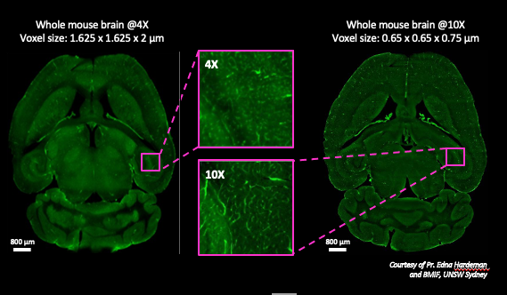 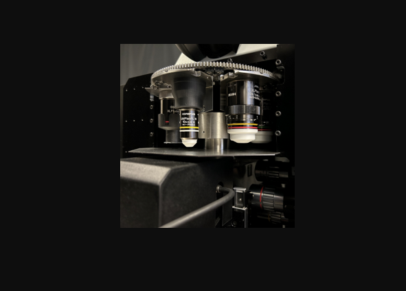 |
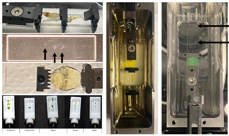 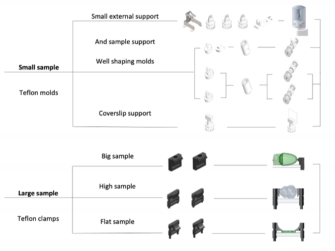 | Easy Multi-Sample MountingThe system provides fast and reproducible mounting of single or multiple samples while preserving the specimen integrity. A wide range of corrosion-resistant sample holders offer the flexibility to image small to large specimens, including cell cultures or whole cleared tissues. For in vivo imaging of live specimens, precise temperature, CO2, and humidity controls are available to maintain stable experimental conditions. |
3D viewer with interactive volume scan settingsThe LINDA 3D viewer generates a quick preview of the sample with a low magnification objective during imaging. This assistant provides a live display of the current slice and builds a 3D render while you are exploring the sample. Intuitive acquisition softwareThe QtSPIM software for the Alpha3 Facility Edition provides intuitive controls for Z-stack, dynamic focus, XY tiling, imaging sequence, focus sweeping, and time-lapse acquisitions. You can select all acquisition parameters, including the laser, camera settings, position of the light sheet, and number of image planes and Z-steps for collecting image datasets with optimal lateral and axial resolutions. | Related Videos |
Need assistance? |
Application Gallery
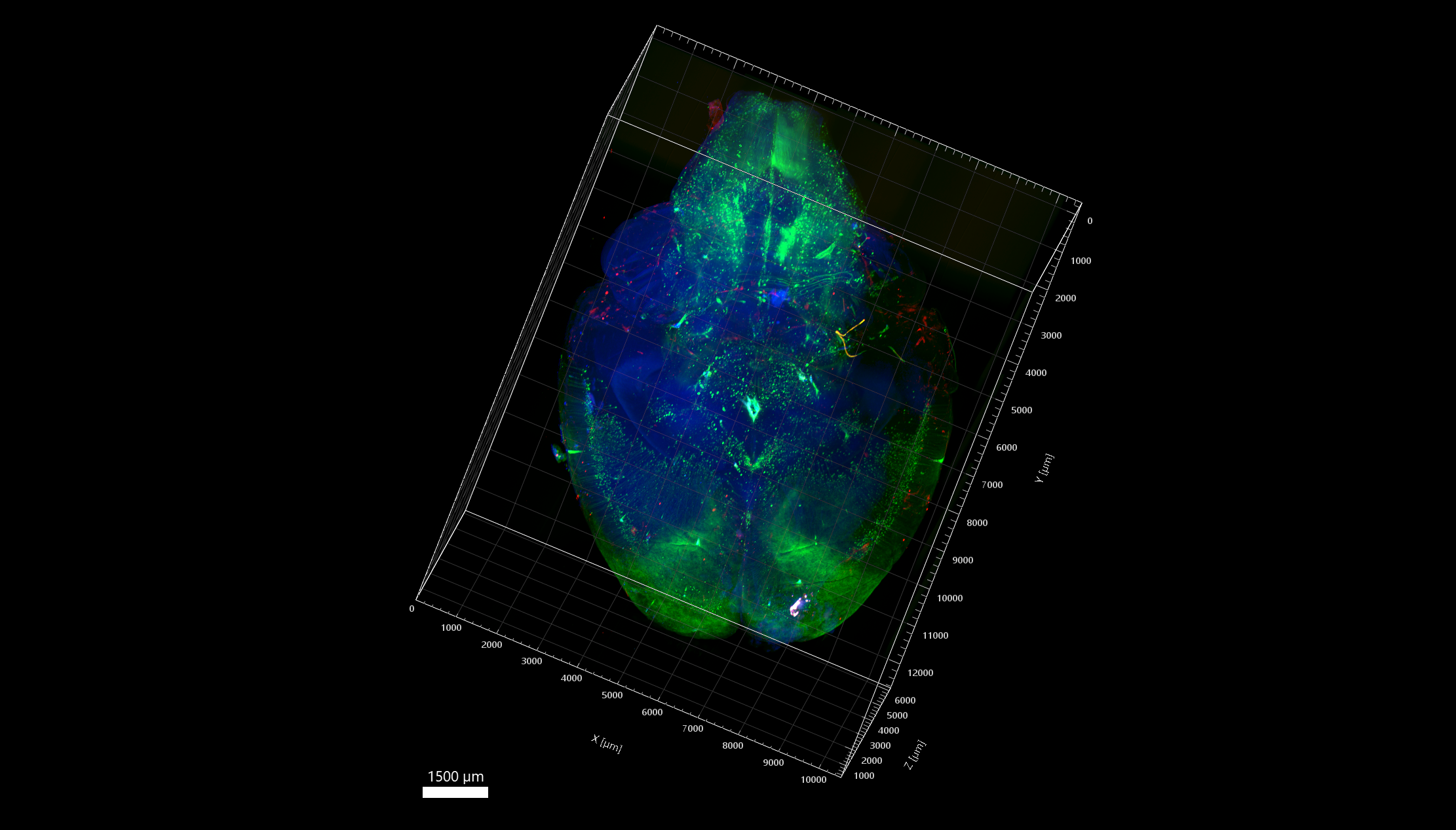 iDISCO cleared adult mouse brain with nuclear GFP staining and cytosolic RFP staining. Captured using an Olympus XLFLUOR4X-340 objective lens (4X/0.28 NA/30 WD). Courtesy of Haohao Wu, Friedrich Miescher Institute for Biomedical Research, University of Basel. | |
Related VideosHuman gastric organoids in PBS1X, with nuclear DAPI staining and membrane CDH1/Alexa 546 staining. | 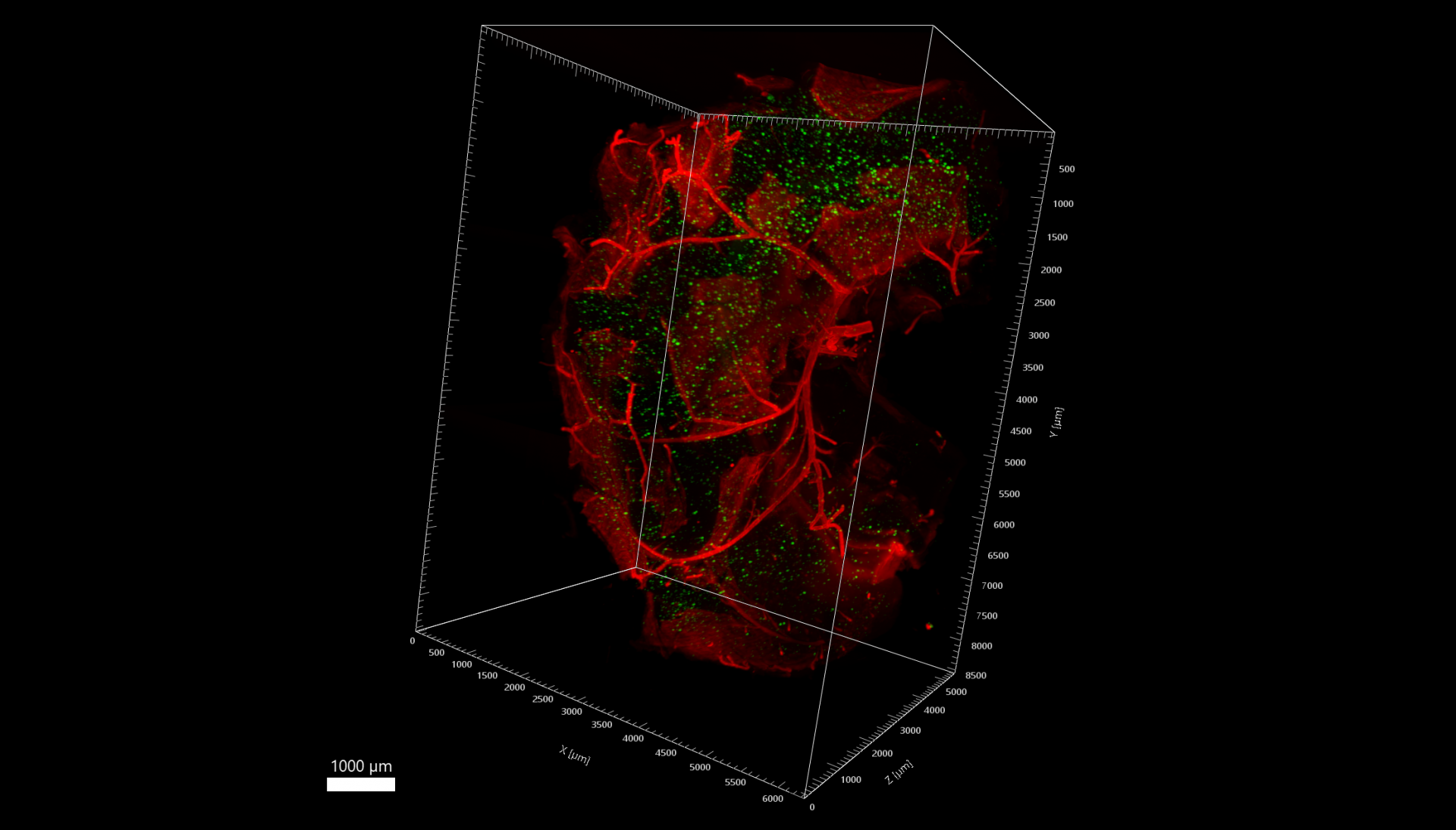 Captured using an Olympus XLFLUOR4X-340 objective lens (4X/0.28 NA/30 WD). Courtesy of Nóra Henn-Mike, Institute of Physiology, Medical School, University of Pécs. |
Related VideosRetinal whole-mount from a Thy1-GCaMP3 mouse. | 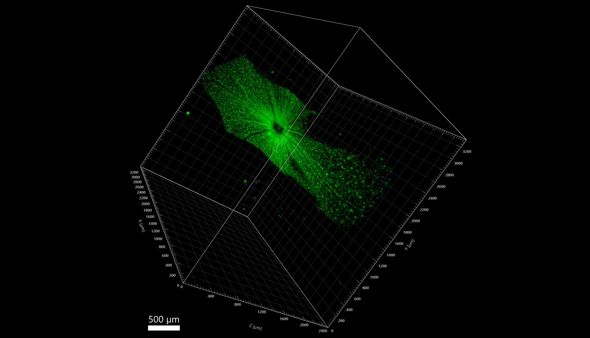 Captured using an Olympus XLFLUOR4X-340 objective lens (4X/0.28 NA/30 WD). Courtesy of Tamas Kovács-Öller, Szentágothai Research Centre. |
Related VideosCUBIC-R+ cleared whole zebrafish larva, YOYO-1 nuclear staining, DiD lipophilic staining, Alexa 647 anti-TH dopaminergic neurons staining. | 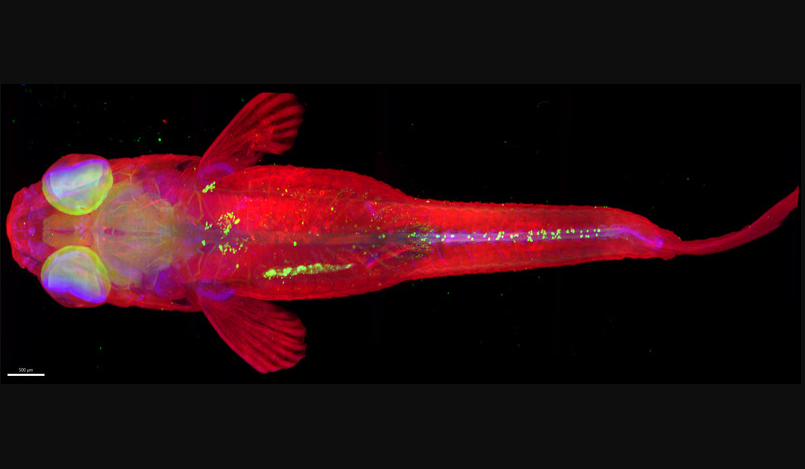 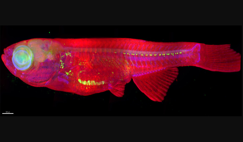 Captured using an Olympus XLFLUOR4X-340 objective lens (4X/0.28 NA/30 WD). Courtesy of Matthieu Simion, TEFOR Paris-Saclay. |