Neuroscience
Life Science Research Application

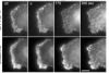
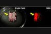
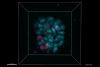
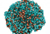
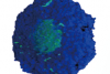
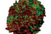
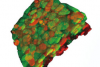
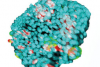
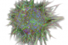
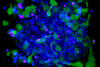
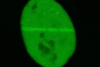
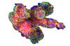
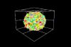
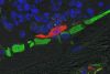
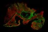
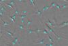
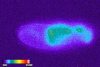
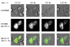
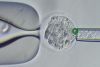
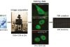
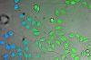
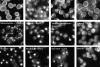
Neuroscience is the study of nerve function, structure, and development. Researchers need to observe single cells as well as the entire brain, on the surface and deep within the tissue. They also need to capture morphological changes and stimulus responses using living samples.
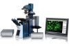
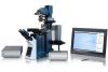
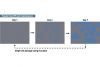
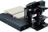
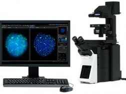
Olympus has a range of microscope solutions to meet the needs of neuroscience research. Our virtual slide scanner system offers the flexibility to view high-resolution digital imaging of the entire slide glass specimen at the same time as the magnified images. The FLUOVIEW confocal laser scanning microscopes enable continuous high-resolution multicolor fluorescence imaging and macro-to-micro observation through highly accurate image lamination. The spinning disk system is designed for live cell imaging with its confocal super resolution microscope, providing high-speed imaging with reduced phototoxicity. Our confocal microscopes capture the fine structure and morphological change of neurons and the deep structure of tissues with super resolution and clear three-dimensional images. They are also capable of capturing the high-speed responses of membrane potential and calcium sensitive dyes. Olympus also offers multiphoton laser scanning microscopes that can capture high-speed reactions and observe deep tissue.
Related Applications (82)


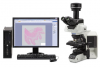


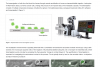

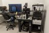

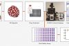


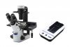

Solutions for Neuroscience - Choose Your ConfigurationMicroscope System/Frame 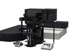 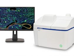 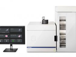 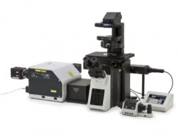  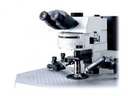 CamerasSoftwareCell Culture Equipment |
Sorry, this page is not
available in your country.