How Old Is the Fish?—Using a SLIDEVIEW™ VS200 Research Slide Scanner to Overcome the Challenges of Inspecting Fish Otoliths
Introduction
Fish in the genus Clupea—also known as herrings—played a pivotal role in the history of marine fisheries in the North Atlantic, and early in the 20th century, their study was fundamental to the evolution of fisheries science (Figure 1). These oily fish also have a long history as an important food.
Herring often move in large schools around fishing banks and near the coast, found particularly in shallow, temperate waters of the North Pacific and North Atlantic Oceans—including the Baltic Sea—as well as off the west coast of South America. Three species of Clupea are recognized and provide about 90% of all herrings captured in fisheries.
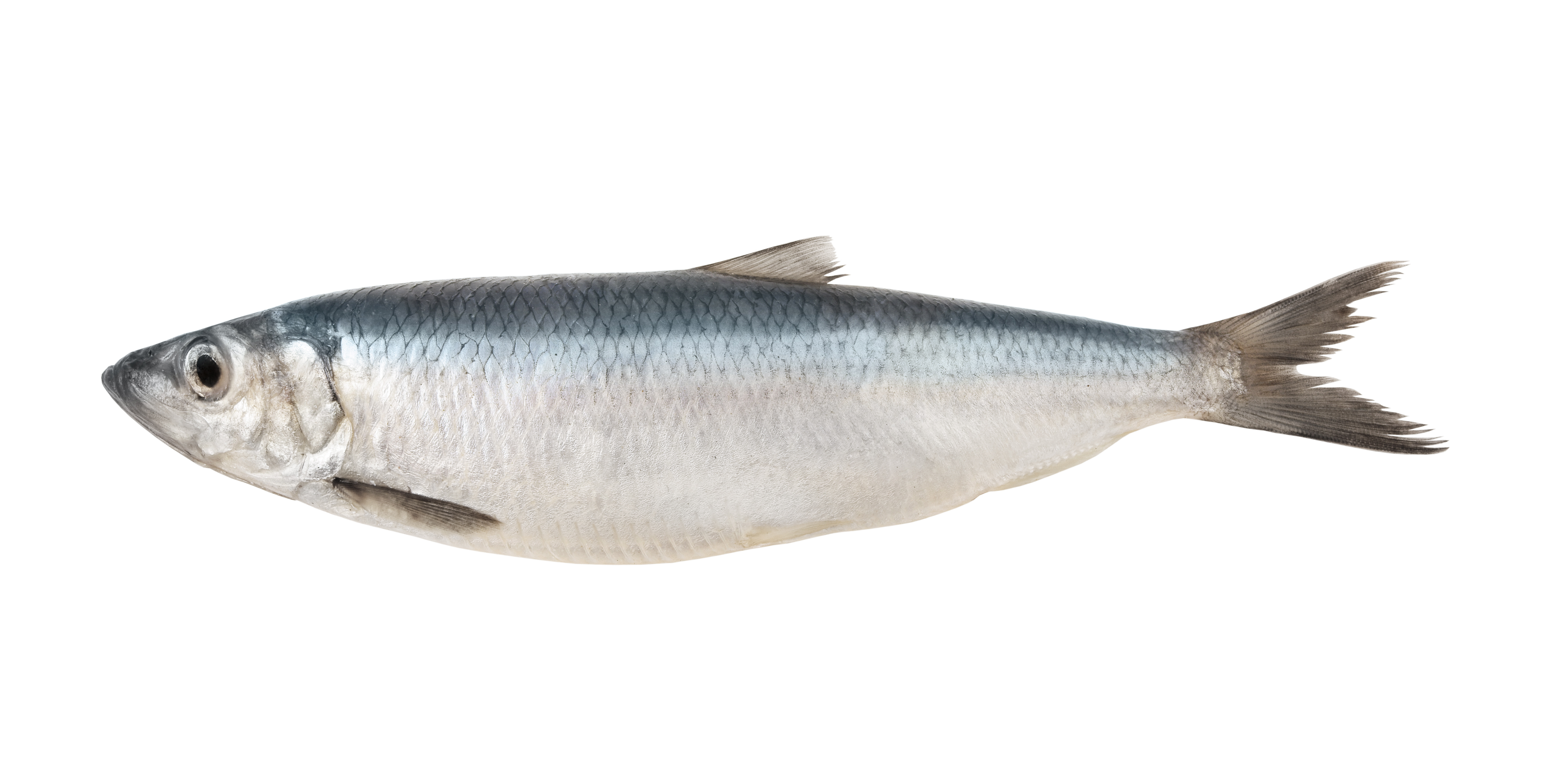
Figure 1. A herring.
Fish age and growth studies are important for understanding the timing and magnitude of spawning, recruitment, habitat use, larval and juvenile duration, and population age structure. Such knowledge is, in turn, important for designing appropriate fisheries management policies. Determining a fish’s age is possible by counting otolith rings.
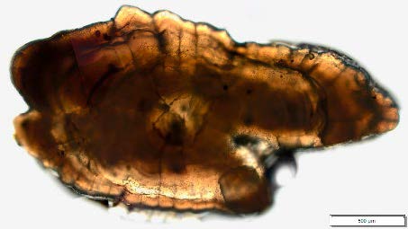
Figure 2. A Herring otolith.
Fish otoliths accrete layers of calcium carbonate and gelatinous matrix throughout a fish’s life. The accretion rate varies with the fish’s growth, typically with less growth in the winter and more in the summer, which results in the appearance of rings that resemble tree rings. By counting the rings, it is possible to determine the fish’s age in years. In addition, in most species the accretion of calcium carbonate and gelatinous matrix alternates in a daily cycle, making it possible to determine a fish’s age in days. This information must be obtained using a microscope and provides important data to early life history studies.
Experiment
Researchers extracted otoliths, embedded them in acrylic resin, and ground them down to a thickness of approximately 50 μm.
Traditionally, a manual upright microscope equipped with polarized light capabilities was used to view the otolith and count the daily rings.
The visibility of the individual layers (rings) changes with different foci and makes this method time-consuming as the observer must constantly refocus the otolith.
The Olympus SLIDEVIEW VS200 slide scanner can be used to automate otolith image capture, saving a significant amount of time (Figure 3). The system can be configured to acquire a Z-stack with up to 31 planes at different magnifications with either polarized light or brightfield. In transmission brightfield, the motorized aperture diaphragm can be adjusted to improve the contrast by closing the aperture in relation to the objective’s numerical aperture. (Figure 4A)
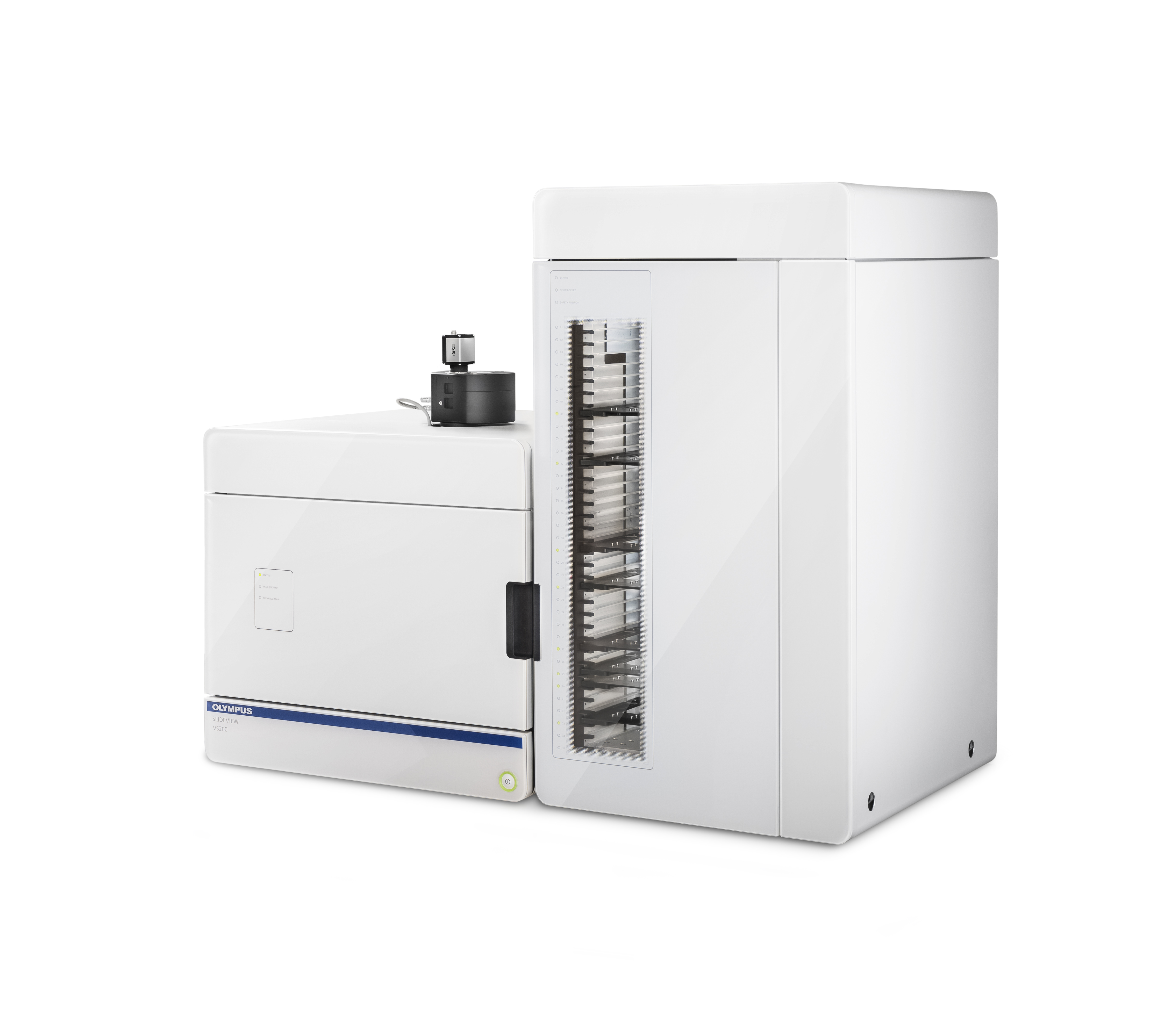
Figure 3. The Olympus SLIDEVIEW VS200 research slide scanner.
Results
Figure 4A. Image of a juvenile herring otolith acquired using a VS200 scanner at 40x (0.95 NA) with an aperture diaphragm opening of 50% as a Z-stack with 18 planes. |
Figure 4B. Adult herring otolith acquired at 40x (0.95 NA) with polarized light as a Z-stack with 30 planes. |
Images courtesy of Dr. Bastian Huwer, National Institute of Aquatic Resources, Section for Marine Living Resources, Lyngby, Denmark |
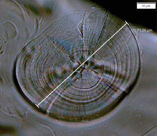
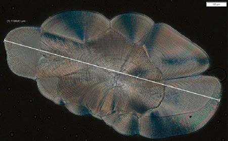
The juvenile otolith has a diameter of approximately 75 μm. The adult otolith has a diameter of approximately 1180 μm. Image 4b was captured using polarized light microscopy, which is designed to observe specimens that are visible primarily due to their optically anisotropic character. The scanner must be equipped with a polarizer positioned in the light path before the specimen and an analyzer (second polarizer) in the optical pathway between the objective rear aperture and the camera port. Image contrast comes from the interaction of plane-polarized light with a birefringent (or doubly refracting) specimen to produce two individual wave components that are each polarized in mutually perpendicular planes.
When concentrating on the center of an otolith, the rings are much denser than in the outer part. This phenomenon is clearly visible in Figures 5A and 5B. The images also demonstrate the importance of acquiring images in multiple Z-planes (a Z-stack) since the rings’ visibility changes at different levels.
The VS200 system automates otolith image acquisition, relieving users of the tedious task of manual focusing and acquisition. Since the images are digitized, it is possible to create a fully automated digital image analysis program to count the otolith layers and measure the age.
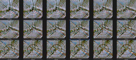 Figure 5A. A cutout of the center of the juvenile otolith Z-stack. Individual Z-planes are shown according to their Z position in μm (Starting from the top left going to the bottom right). The distance between each Z-plane is 1.17 μm. The Z-stack spans a distance of 20 μm, and the distance between the otolith layers in the center part is only about 600 nm. | 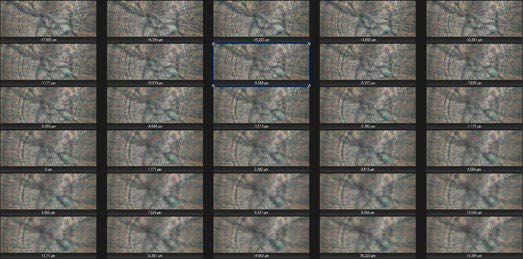 Figure 5B. A cutout of the center of the adult otolith Z-stack. Individual Z-planes are shown according to their Z position in μm. The distance between each Z-plane is 1.17 μm. The Z-stack spans a distance of 34 μm, and the distance between the otolith layers in the center part is about 740 nm. |
Conclusion
The SLIDEVIEW VS200 research slide scanner offers an easy, convenient way to acquire images of resin embedded fish otoliths. In juvenile fish, the otolith layer distance is only around 600 nm, requiring an imaging system capable of high magnification and resolution. Using a 40x UPlanXApo objective with an NA of 0.95, the VS200 system offers the magnification and resolution required to capture these images.
Adult otolith specimens are also quite thick (up to 34 μm) and not very transparent since they are made from calcium carbonate. The VS200 system has advanced features that can overcome these challenges and obtain high-quality images. For thick samples, the system can scan the sample at multiple Z-planes with polarized light. To improve contrast in otolith samples, the system’s aperture diaphragm can be closed to make the age rings more visible for easy counting.
Acknowledgments
This application note was prepared with the collaboration of Dr. Bastian Huwer, National Institute of Aquatic Resources, Section for Marine Living Resources, Lyngby, Denmark
Products related to this application
Sorry, this page is not
available in your country.