Rapid Automated Detection and Segmentation of Glomeruli Using Self-Learning AI Technology
Researchers often rely on analyzing large image sets to obtain relevant data for their investigations. These analyses can be both quantitative and qualitative, and they must also be reliable and unbiased. Depending on the morphological area of interest, this task can be quite cumbersome. To accelerate this process, Olympus offers a deep-learning solution in combination with the SLIDEVIEW™ VS200 scanner. In this experiment, the scanner’s self-learning image analysis software module was applied to the detection of glomeruli in the kidney.
Kidney Glomeruli Detection and Segmentation
In renal pathology research, scientists work to unravel the complex processes of diseases associated with the kidney to be able to develop better, more efficient treatments.
Kidneys perform many vital functions that help maintain our overall health with the help of glomeruli. Glomeruli are renal structures that filter plasma and maintain homeostasis. Glomeruli can be affected in several disease conditions, which are being extensively studied by scientists. An essential part of this scientific research involves analyzing a significant number of kidney sections, which are prepared using a wide variety of staining protocols to reveal these specific structures.
Analysis of images of kidney sections can be crucial in determining changes in glomerular morphology or numbers. However, processing multiple samples for research can be time consuming and subjective. To accelerate and optimize this process, nephropathology researchers can use the SLIDEVIEW VS200 digital slide scanning system to rapidly scan huge numbers of slides. The system’s image analysis software now has a deep-learning neural network module that can be trained to recognize glomeruli regardless of variations in size, shape, or color.
Challenges of Automatically Detecting and Segmenting Glomeruli
The complexity of renal tissue, the heterogeneity of the pathological features, and the conventional use of multiple stains for renal histology preparations make computer-aided image analysis of renal biopsy samples challenging. Staining tissue sections can also give each slide different color intensities, depending on the amount of stain present.
All this variability related to the glomerular appearance, and to digital tissue images in general, makes it difficult to automate the classification. This is why the application of glomeruli detection in kidney tissue is a particularly rigorous test for the viability and efficacy of the VS200 slide scanner and TruAI™ deep-learning solution.
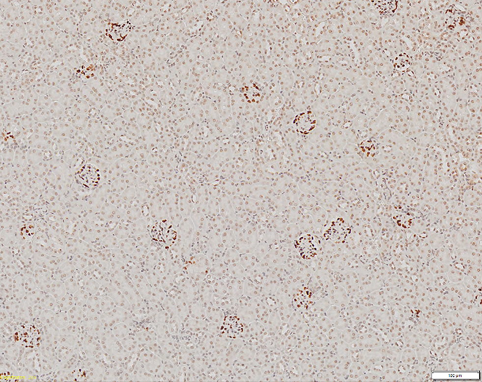 | 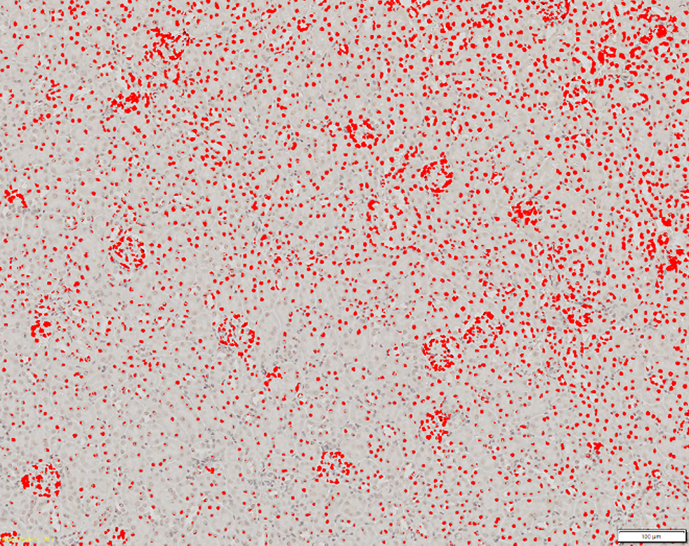 |
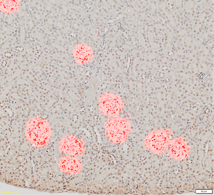
Slide (left) of a kidney tissue section at 20x with glomeruli stained in dark brown and (right) at 10x showing the detection based on the conventional threshold method (in red) failing to distinguish the glomeruli cells from other tissue cells. |
Experiment Using the SLIDEVIEW™ VS200 Scanner and TruAI™ Deep-Learning Solution
To analyze these structures in scanned images, normally researchers select the glomeruli manually, which is highly time consuming. Moreover, traditional automatic segmentation methods, such as threshold-based algorithms, fail to exclusively detect them, as shown above.
The VS200 slide scanning package includes the VS-Desktop software for image analysis. This software offers a TruAI™ module, which applies a deep-learning approach to detection and segmentation based on convolutional neural networks. These networks are a form of self-learning microscopy and are an extremely powerful technology for object segmentation. Thanks to this technology, we can automate the detection of the glomeruli in a kidney.
The neural network training process is explained in the next section.
Easy Workflow to Automatically Detect and Segment Samples Using Deep LearningThe first step toward this automatic analysis is to provide the software with annotated sample images or “ground truth” data. This is created by hand labeling the glomeruli in a small number of images in the VS-Desktop software. The more objects that are marked, the better. Higher numbers of objects with differences in intensity, color, size, and shape make the neural network more robust. The VS image analysis software has various tools to facilitate this step, making it easy to use. | 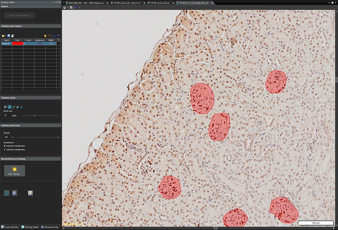 | |
 | 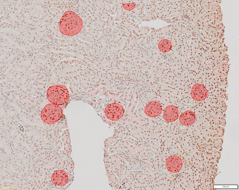 | 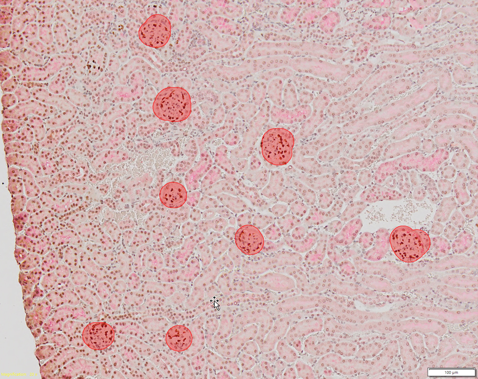 |
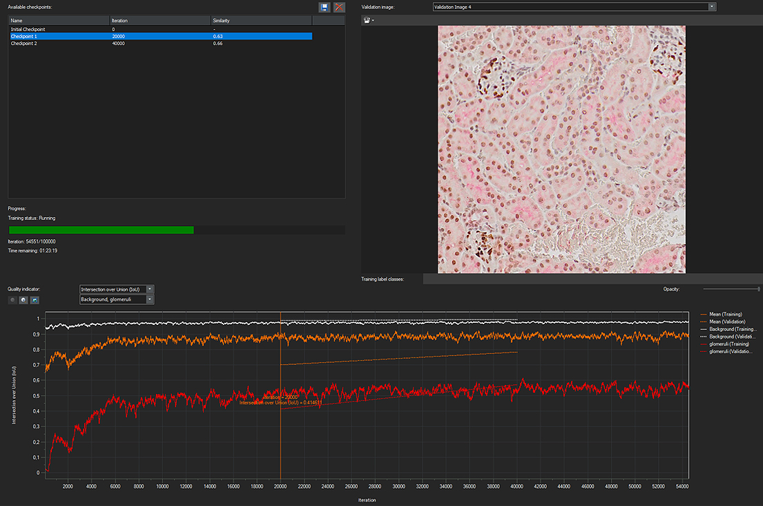
After the labeled data is generated, the next step is training the deep neural network (DNN). In this phase, the network compares the ground truth data with its own calculated data until a high probability value is reached. This calculated data is a type of artificial intelligence (AI) that imitates the human brain (the so-called DNN), which learns to recognize structures and make intelligent judgements. |
Lastly, the calculated DNN is applied to the rest of the kidney images to automatically detect and separate the glomeruli.
A trained Olympus TruAI™ deep neural network can be transferred and used at any VS200 desktop station as well as other compatible Olympus products.
This process results in an automated workflow as shown below. Segmenting glomeruli includes these three simple steps:
1. A new image is scanned with the SLIDEVIEW™ VS200 digital slide scanning system.
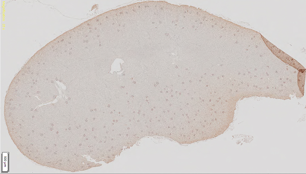 |
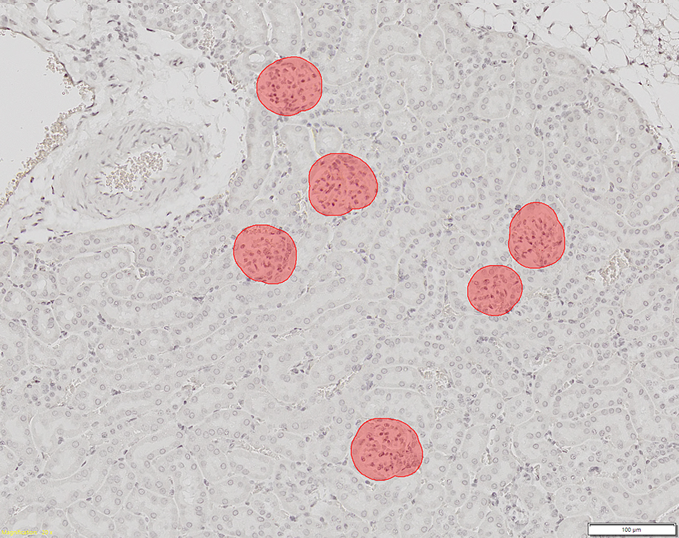
2. Glomeruli are detected and separated by the trained DNN.
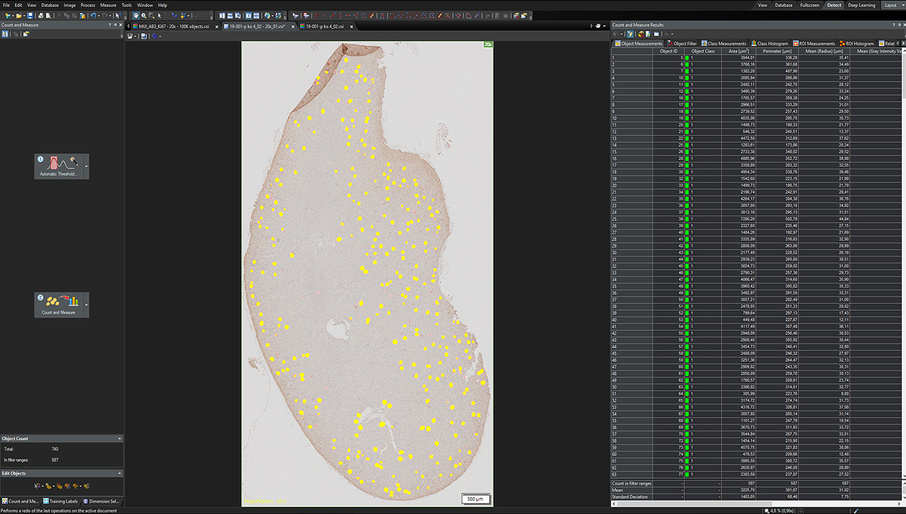
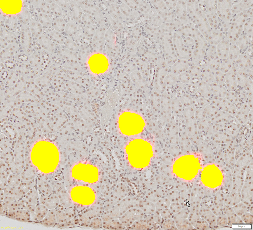
3. The detected glomeruli can be segmented and used to perform further Count and Measure analysis on the output image.
 |  |
Summary of the Advantages Provided by the SLIDEVIEW VS200 Scanner with Deep-Learning Solution for the Detection and Segmentation of Glomeruli
The TruAI™ module enabled easy object detection and segmentation of glomeruli from complex images with much higher reliability and accuracy than other existing automatic methods. Further analysis, such as counting and measurement, can also be performed based on these segmentation results.
The combination of the SLIDEVIEW™ VS200 scanner and TruAI™ deep-learning solution can provide a complete workflow from sample acquisition to accurate quantitative data analysis in a wide range of biological applications on a variety of images, such as cells and tissue samples in brightfield and fluorescence.
Precise automation of image analysis will relieve a lot of the tedious manual work for scientists, making their research more efficient.
Authors
- Zhenhua Miao, Principal Scientist at ChemoCentryx, Inc. Mountain View, CA, USA
- Sara Quiñones Gonzalez, Product Manager, Olympus Soft Imaging Solutions GmbH, Münster, Germany
- Dr. Daniel Göttel, Sales Support Manager, Olympus Soft Imaging Solutions, Münster, Germany
Products Related to This Application
Sorry, this page is not
available in your country.