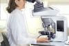
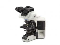
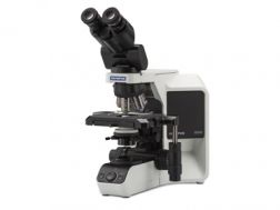
In cytology and pathology research, specimens taken from cells, tissues, or organs are tested with the naked eye or using a microscope to study the changes that occur when they are affected by disease. We provide solutions particularly suited for pathological research applications through specialized microscope, camera, and imaging software systems. Our line of upright microscopes offers high optical performance with true color reproducibility. Ergonomically designed, they help ensure
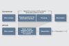
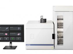
comfortable observation over long periods. Our robust slide loader system is designed for research and pathology, offering simultaneous viewing of the whole slide and the magnified region and an innovative synchronizing function that can be used to analyze multiple virtual slides. This innovative virtual slide system for high-resolution digital imaging also enables you to easily view the entire slide glass specimen. Specimen preservation, database construction, next-generation
education/training, and consultation are also offered for this field of research.
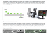
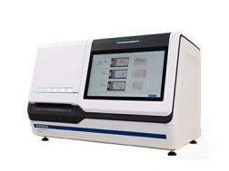
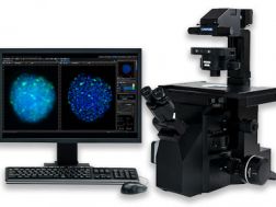
The APEXVIEW™ APX100 benchtop fluorescence microscope makes it fast and simple to acquire expert-quality microscope images. Built with our renowned optics, an intuitive user interface, a powerful AI, and a suite of smart features, the APX100 system combines ease of use with high-quality image data to fit your research needs.
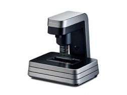
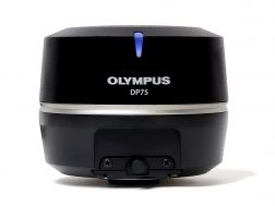
The high-performance DP75 digital microscope camera makes it easy to capture high-resolution brightfield or fluorescence images using a single color camera. It simplifies your microscopy imaging, so you can focus more on your work.
Integrated TruAI denoising maximizes the camera’s image quality in real time
Exceptional color reproduction, making your images as vivid as looking through the microscope oculars
Supports multiple staining combinations and wavelengths up to 1000 nm with a switchable infrared (IR) cut filter
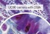
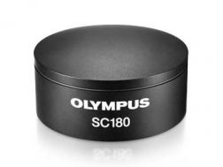
See and document a sample’s fine details and structures, using the high-resolution SC180 microscope digital camera. Equipped with an 18-megapixel color CMOS sensor, this camera provides fast 4K UHD live imaging displayed at 25 fps.
18-megapixel color CMOS sensor to document fine details
Vivid, low-noise images for insightful observations
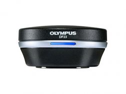
Enabling fast, easy capture of high-quality images that can be clearly observed on a large screen, the DP23 microscope digital camera eases routine life science and clinical research, conferencing, or teaching. Integrate it seamlessly into your microscopy workflow and easily share or stream images.
Share images using the DP23-AOU network solution
Clearly observe live images on a large screen
Fast, high-quality imaging for conferences and teaching
Providing color accuracy and 4K resolution, the DP28 digital microscope camera’s powerful features and wide field of view capture images that enhance tasks such as conferencing, teaching, and clinical research. Integrate it seamlessly into your microscopy workflow for improved work efficiency and image quality.
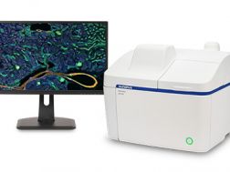
Providing intuitive operations and a seamless workflow, cellSens software’s user interface is customizable so you control the layout. Offered in a range of packages, cellSens software provides a variety of features optimized for your specific imaging needs. Its Graphic Experiment Manager and Well Navigator features facilitate 5D image acquisition. Achieve improved resolution through TruSight™ deconvolution and share your images using Conference Mode.
Improve experiment efficiency with TruAI™ deep-learning segmentation analysis, providing label-free nuclei detection and cell counting
Modular imaging software platform
Intuitive application-driven user interface
Broad feature set, ranging from simple snapshot to advanced multidimensional real-time experiments