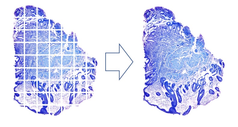
Create high-resolution, whole-slide images in real time simply by moving the controls of a manual stage with the Instant Multiple Image Alignment (MIA) function.
High-resolution images suitable for teleconsultation or education are easily achieved with this cost-effective solution.
Bright, sharp images: Our quality optics combined with True Color LED technology ensure that you are seeing everything your samples have to offer.
Easy collaboration: Conference Mode enables you to share images with just one click and easily mark them up.
Faithfully reproduce colors: Cameras, like the DP74, with proprietary image processing technology detect subtle shading and deliver excellent color fidelity in a wide variety of stained samples.
Telepathology: Our NetCam Solution uses standard TCP/IP protocols to facilitate live viewing for teaching, mentoring, or supervision in and outside the lab. Our solution can also be used with general web meeting software.
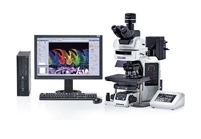
Standalone system: miniPCs or tabletPCs enable small footprint imaging system with our cellSens
Real-Time Panoramic Imaging with cellSens® Instant MIA
Create high-resolution, whole-slide images in real time simply by moving the controls of a manual stage with the Instant Multiple Image Alignment (MIA) function.
Panoramic images, fast: Real-time image stitching with the Manual Process solution.*
No motorized stage required: Use a manual stage to create seamless panoramic images with smooth control over the XY stage controls.
Fully automated panoramic imaging: A motorized stage and cellSens Dimension software with the Multiposition solution enable automated whole-slide image acquisition. Add a motorized Z correct for the effects of sample distortion and tilting.
Minimize stitching errors: Correct for common problems associated with image stitching with cellSens software; image calibrations are automatically applied to live images.
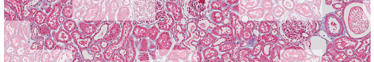
Discover the Detail without Losing the Big Picture
Seamless wide-area images of whole tissue sections are generated from multiple images acquired in high magnification and corrected for small mismatches, enabling you to visualize the entire tissue section. Focus view makes it easy to correct for differences in tissue thickness as the image is acquired, eliminating the need for time-intensive Z-stacks.
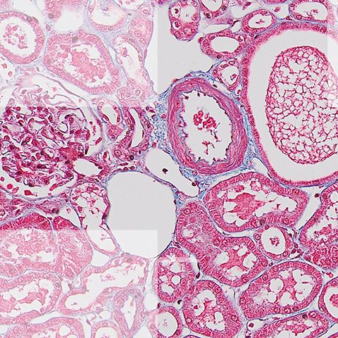
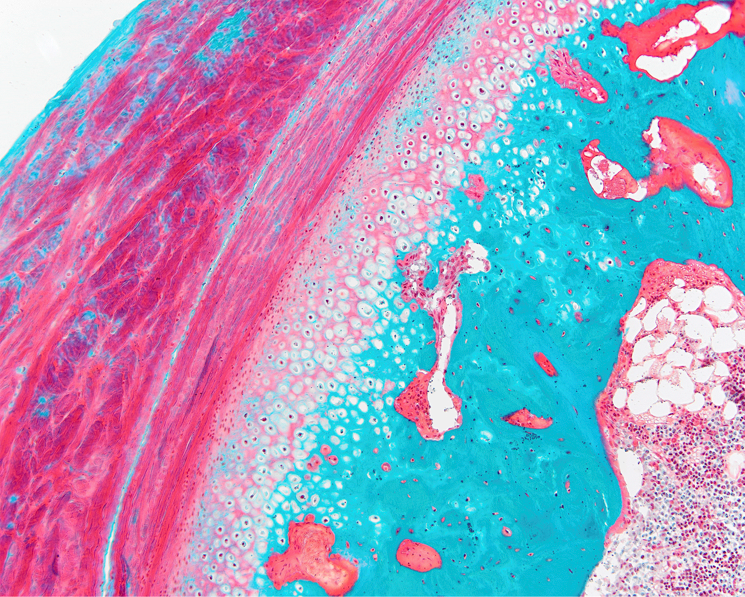
Bone Section | 10x Magnification
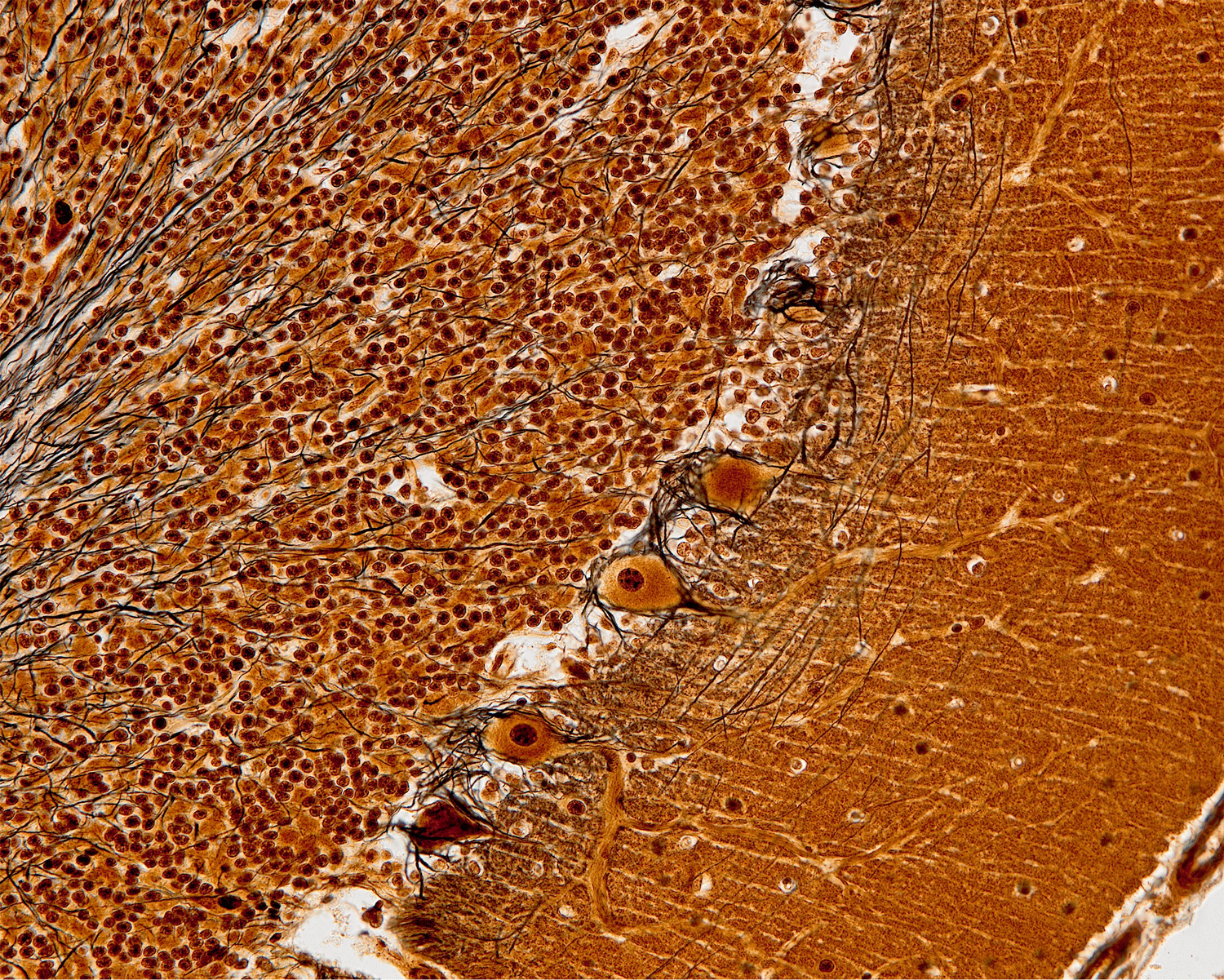
Cerebellum Section | 10x Magnification
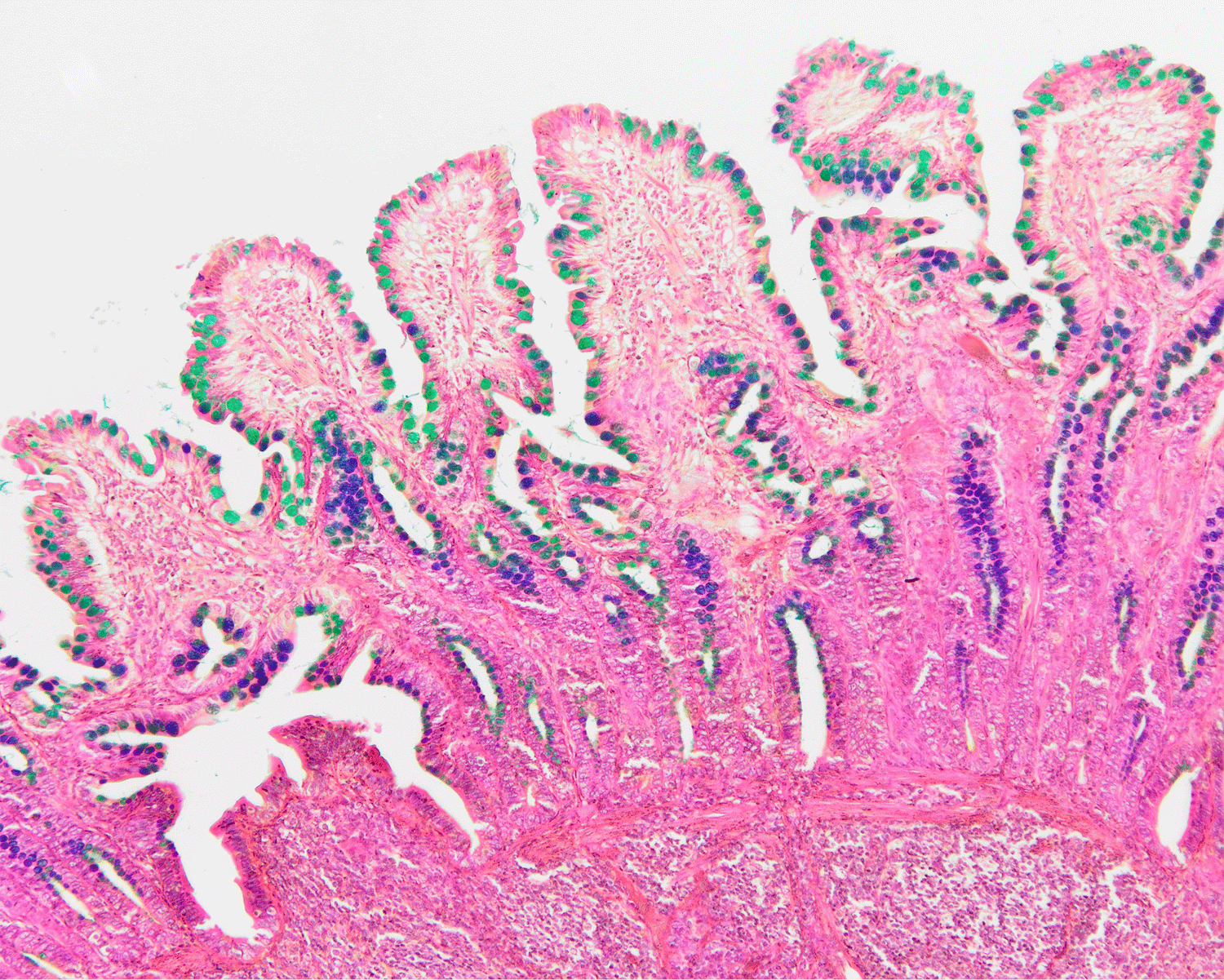
Intestine Section | 10x Magnification
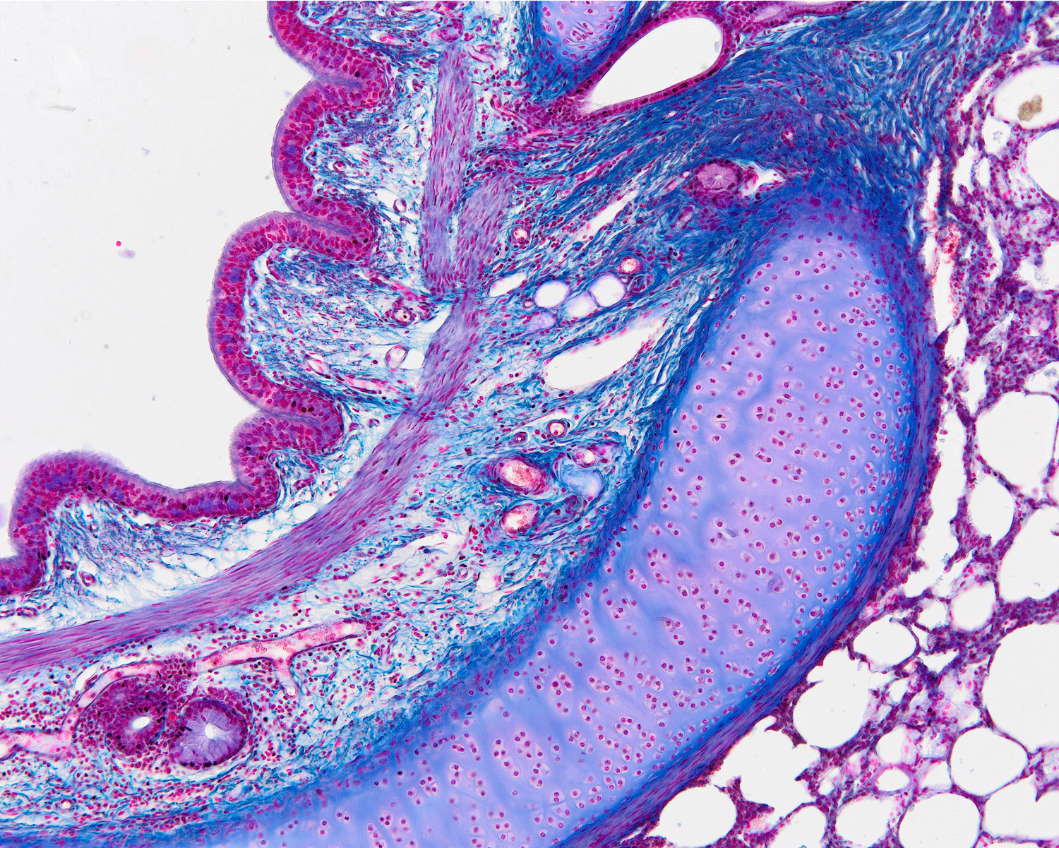
Lung Section | 10x Magnification
Microscopy in 4K
The 9-megapixel UC90 camera from Olympus captures it all. Whatever your imaging needs are, expect no less than exceptional results in image quality, sensitivity, dynamic range, and color fidelity. The 4K UHD Live Image from the Olympus UC90 camera fully utilizes modern 4K display capabilities, making it an ideal solution when the details matter.
Involve every participant: Deliver enhanced on-screen discussions without the need to share the oculars.
Simple navigation: Switch between different observation modes with a single click.
Locate regions of interest: High frame rate and fluid sample navigation and focusing make it easy to find regions of interest.
Noise reduction: OISA technology enables viewing without the added noise traditionally associated with digital microscope cameras.
Applicable Products
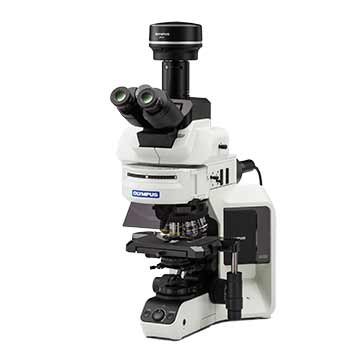
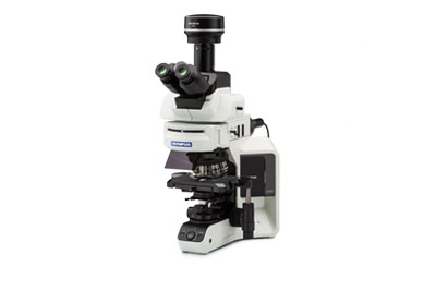
BX53 Upright Microscope
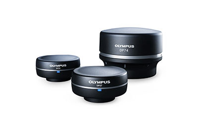
Digital Cameras
cellSens Imaging Software
*Manual Process solution available as an option for cellSens Entry and Standard software and is included within cellSens Dimension software.
Not Available in Your Country
Sorry, this page is not
available in your country.
Not Available in Your Country
Sorry, this page is not
available in your country.