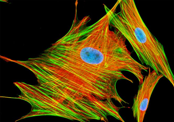
Mongoose Skin Fibroblast Cells (APM)
Immunofluorescence with mouse anti-alpha-tubulin was employed to visualize distribution of the microtubule network in a log phase monolayer culture of African water mongoose skin cells. The secondary antibody (goat anti-mouse IgG) was conjugated to Alexa Fluor 568 and mixed with Alexa Fluor 488 conjugated to phalloidin to simultaneously image tubulin and the actin cytoskeleton. Nuclei were counterstained with Hoechst 33258.
Sorry, this page is not
available in your country.