
New Year’s Eve may have been a couple months ago, but the top images for this month look ready to party! From previous Image of the Year winners to aquatic microorganisms, your favorite images for February are bright and bold.
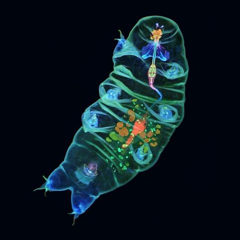
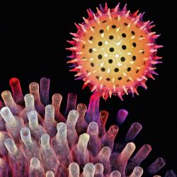
Our top image of the month is a reappearance of our Image of the Year 2019 regional winner for the Americas. This adorable tardigrade looks like it’s greeting good friends. According to Tagide deCarvalho, it may just be ready for a snack.
“This is an image of a live tardigrade, also known as a water bear or moss piglet, where you can clearly see the digestive tract from the mouth all the way down to the cloaca. At the front end, you can see that the mouth is a tube armed with stylets used to pierce food, such as plant cells or algae, and the muscular pharynx is used to suck in the juices.”
Image and caption courtesy of Tagide deCarvalho, the Image of the Year 2019 regional winner for the Americas. To learn more about Tagide’s winning image, read our interview.
To enter this year’s Image of the Year competition for a chance to win your own microscope or objectives, visit olympus-lifescience.com/ioty.
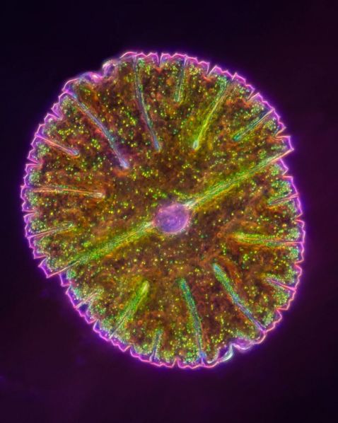
Science or poetry? We see both in this shimmering sample.
“Like a delicate jewel suspended in a miniature universe, the cell body of the Micrasterias desmid is adorned with intricate patterns, with internal crystals that shimmer and dance as they are pulled along by motor proteins.” A unicellular green alga, Micrasterias displays a beautiful symmetry.
Image and caption courtesy of Karl Gaff. Captured using a BX51 microscope at 400X magnification.
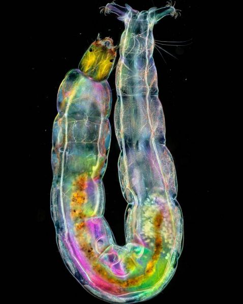
Where’s the nearest dance floor? This midge larva looks ready to meet the tardigrade and head to the disco!
“Revealed through darkfield polarization microscopy with a BX51 microscope, this captivating image showcases the radiant hues of a midge fly's aquatic phase. It's tree-like nervous system and herbivorous diet become apparent, portrayed in a panoramic composition achieved through meticulous manual focus stacking of five stacks, each consisting of up to 80 images.”
Image and caption courtesy of Karl Gaff. Captured using a BX51 microscope with an X Line™ 20X objective.
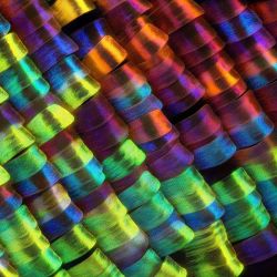

We always have a good time celebrating our past Image of the Year winners! Appearing in our top image roundup are the three regional winners of the 2022 competition. From left to right:
- Scales of the wing of the Urania rhipheus moth. Captured at 20X by Javier Ruperez (Spain).
- Depth color-coded projection showing a germinating pollen grain of a morning glory attached to the stigma. Captured by Igor Siwanowicz (USA).
- Edelweiss stamens, which were scanned and reconstructed in three dimensions using confocal laser scanning microscopy. Captured by Jiao Li (China).
To view past winners and download our desktop and phone backgrounds, visit olympus-lifescience.com/ioty.
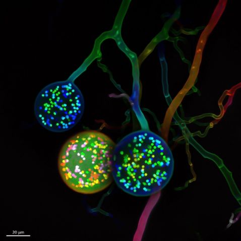
Believe it or not, these bright bulbs aren’t party lights! These glowing orbs are actually the multinucleate spores of a soil fungus.
“The image depicts three spores (circular formations) of a particular fungal group known as arbuscular mycorrhizal (AM) fungi. Every colorful bright spot within the spores corresponds to a single nucleus. The more abstract lines behind the spores depict the filamentous body of the fungus. These structures are known as hyphae, and they are similar to open pipes where nutrients, water, and organelles travel with impressive speed at long distances.”
Image and caption courtesy of Vasilis Kokkoris, the Image of the Year 2021 regional winner for EMEA. To learn more about Vasilis’ winning image, read our interview.
Bonus video! Bringing a party of organisms to the microscope looks like fun.
“Here’s a short compilation of organisms I found all around town—from the cemetery pond I go to every week to my own stock tank I keep in the backyard (most people find that to be some weird science).
Clips 1–3 are ciliates and the last two are rotifers. That very last one is my fave!”
Video and caption courtesy of Desi Morrison. Captured using a BX51 microscope.
Share Your Best Light Microscopy Images
To see more images like these, be sure to follow us on Instagram at @evidentlifescience!
Interested in sharing your own images? Enter this year’s Image of the Year competition before time runs out! Upload your best light microscopy images (up to three images and three video files) at olympus-lifescience.com/ioty.
You can also share your own microscope images through our image submission site!
Related Content
Cells to Crystals—Our Most Popular Microscope Images for January 2024
Stars and Smiles—Our Most Popular Microscope Images for December 2023
Over the Rainbow—Our Most Popular Microscope Images for November 2023
