Evident LogoOlympus Logo

Ask the Experts

Metabolic Imaging in Langerhans Human Islets with MPE and FLIM

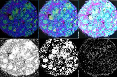
Capturing life (mis)regulation at the nanoscale is a crucial challenge for present and future biophysics. At this scale, the main actors are the molecules. To successfully tackle molecular behavior within living matter, optical microscopy is a valuable methodological platform: by using fluorescence as the signal, spatial and temporal details of molecular processes can be investigated quantitatively. The physiopathology of beta-cell response to glucose stimulation will be used as case study of biological/biomedical interest. The metabolic traits of beta cells will be highlighted using a straightforward combination of multiphoton microscopy, fluorescence lifetime imaging, and feedback-based orbital tracking of sub-cellular nanostructures.

Presenter: Francesco Cardarelli
Associate Professor in Applied Physics, Scuola Normale Superiore of Pisa

After receiving his M.Sc. in Biological Sciences from the University of Pisa in Oct 2003 and his Diploma in Biological Sciences in the same year (both with honors) from SNS, Francesco Cardarelli worked at the NEST Laboratory of SNS as a PhD student in Molecular Biophysics under the supervision of Prof. Fabio Beltram. He started his interdisciplinary research at the crossroads between cell biology and physics, using advanced fluorescence microscopy methods to study the intracellular transport properties of virus-derived peptide sequences. After graduating, he became a Post-Doctoral Fellow at the Laboratory for Fluorescence Dynamics, University of California at Irvine, under the supervision of Prof. Enrico Gratton, where he coordinated the research activity for the development of new spatial variants of fluorescence correlation spectroscopy to detect barriers to molecular diffusion/flow in live cells. In Dec 2010 he was hired by the CNI@NEST (IIT) as a Post-Doctoral Fellow. Back in Italy, he started working to develop new fluorescence-based imaging and analysis methods to study single molecules in complex biological systems with high spatiotemporal resolution. This research was boosted by a number of funded grants (and established collaborations) and by an independent scientific position, first at CNR as a Researcher, then at SNS as Professor in Applied Physics.

The focus of his research is on the development of new optical microscopy techniques to increase the amount of quantitative information that can be extracted from investigations on living matter. For instance, in recent years, he and his team introduced a number of new spatiotemporal fluctuation-analysis tools (iMSD, iRICS, nD-pCF, diffusion tensor analysis, etc.) to extract structural and dynamic properties of biological objects, from molecules to entire sub-cellular structures, in their complex natural environment. Such a toolbox is becoming a new paradigm for biophysical investigations at the nanoscale, as featured in the “New and Notable” section of Biophysical Journal (2016 Aug 23; 111(4): 677–678). In 2014, together with his Team, they demonstrated the occurrence of short-range protein Brownian motion in the cell cytoplasm, being among the first to challenge the current view of the structural organization of the crowded intracellular environment. Finally, by combining this toolbox with feedback-based orbital tracking, they demonstrated that even the nanoscopic and dynamic environment of intracellular organelles can be quantitatively probed.

Experten
Francesco Cardarelli
Associate Professor in Applied Physics
Scuola Normale Superiore of Pisa

After receiving his M.Sc. in Biological Sciences from the University of Pisa in Oct 2003 and his Diploma in Biological Sciences in the same year (both with honors) from SNS, Francesco Cardarelli worked at the NEST Laboratory of SNS as a Ph.D. student in Molecular Biophysics under the supervision of Prof. Fabio Beltram. He started his interdisciplinary research at the crossroads between cell biology and physics, using advanced fluorescence microscopy methods to study the intracellular transport properties of virus-derived peptide sequences. After graduating, he became a Post-Doctoral Fellow at the Laboratory for Fluorescence Dynamics, University of California at Irvine, under the supervision of Prof. Enrico Gratton, where he coordinated the research activity for the development of new spatial variants of fluorescence correlation spectroscopy to detect barriers to molecular diffusion/flow in live cells. In Dec 2010 he was hired by the CNI@NEST (IIT) as a Post-Doctoral Fellow. Back in Italy, he started working to develop new fluorescence-based imaging and analysis methods to study single molecules in complex biological systems with high spatiotemporal resolution. This research was boosted by a number of funded grants (and established collaborations) and by an independent scientific position, first at CNR as a Researcher, then at SNS as Professor in Applied Physics.

The focus of his research is on the development of new optical microscopy techniques to increase the amount of quantitative information that can be extracted from investigations on living matter. For instance, in recent years, he and his team introduced a number of new spatiotemporal fluctuation-analysis tools (iMSD, iRICS, nD-pCF, diffusion tensor analysis, etc.) to extract structural and dynamic properties of biological objects, from molecules to entire sub-cellular structures, in their complex natural environment. Such a toolbox is becoming a new paradigm for biophysical investigations at the nanoscale, as featured in the “New and Notable” section of Biophysical Journal (2016 Aug 23; 111(4): 677–678). In 2014, together with his Team, they demonstrated the occurrence of short-range protein Brownian motion in the cell cytoplasm, being among the first to challenge the current view of the structural organization of the crowded intracellular environment. Finally, by combining this toolbox with feedback-based orbital tracking, they demonstrated that even the nanoscopic and dynamic environment of intracellular organelles can be quantitatively probed.

Metabolic Imaging in Langerhans Human Islets with MPE and FLIM7.9.2024
Sorry, this page is not available in your country
Ask the Expert Sign-up

By clicking subscribe you are agreeing to our privacy policy which can be found here.

Sorry, this page is not
available in your country.

Sorry, this page is not available in your country