
From salt and stinging nettles to samples from Sagami Bay, the top five images from May show just how much beauty one cell can hold.
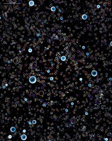
Diatoms are unicellular, but while they consist of one cell, their wide variety of shapes, sizes, and patterns offer countless imaging opportunities under the microscope. The bubble-like diatoms seen here were sampled in a plankton net in Sagami Bay, Japan. These diatoms are from a sample that sank to the bottom of the petri dish.
Image courtesy of @co_micro. Captured using an Olympus BH2 microscope.
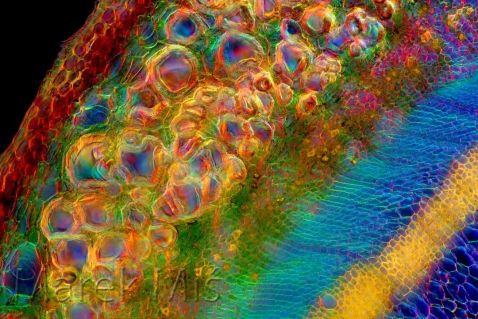
Red and orange, green and blue! These are the colors in a common children’s song about rainbows, as well as some of the vivid colors seen in this stunning transversal cross-section of a nettle (Urtica sp.) stalk. The fresh, unstained material was cut in fingers (without a microtome) with a shaving razor, then imaged through the microscope under polarized light and darkfield.
Image courtesy of Marek Miś. Captured using an Olympus BH2 microscope.
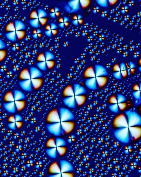
Last month’s top images post included a beautiful red, white, and blue image of Ammonaps, or sodium phenylbutyrate, which we noted is used to treat urea cycle disorders. Once again, this salt compound appears in our roundup, this time showing a selection of the crystals under polarized light.
Image and caption courtesy of Håkan Kvarnström. Captured using an Olympus UPLXAPO 4X objective.
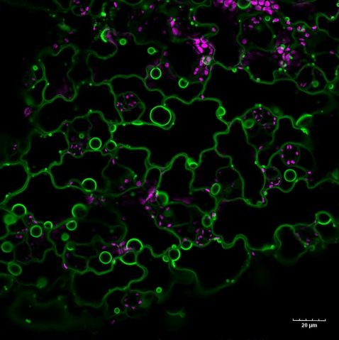
“This is a section of the cotyledon (first leaf) surface from the flowering plant Arabidopsis thaliana, which is expressing the green fluorescent protein vacuolar membrane marker (vac-GB). Chloroplasts are in magenta, visible due to the autofluorescence of the chlorophyll.
Fun fact: in mature plant cells, vacuoles occupy most of the cell volume (>90%). The entire cytosol of two neighboring cells and their shared cell wall are squished into small spaces between two large vacuoles you can see in parts of the image.”
Image and caption part of an Instagram takeover courtesy of Ivan Radin, Americas regional winner of the 2021 Image of the Year competition. Captured using an Olympus FV1000 confocal microscope. To learn more about Ivan, check out our interview here.
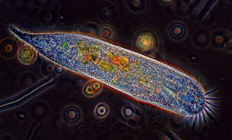
Pictured here is a ciliate imaged using phase contrast, proving yet again that in only one cell you can find a great deal of beauty.
Image courtesy of Marek Miś. Captured using an Olympus BH2 microscope.
Check out this bonus video of an unforgettable tree!
Ginkgo biloba, commonly known as ginkgo or gingko, is one of the oldest living tree species, and it looks mesmerizing under the microscope.
"The arrangement of ginkgo cells in the stem is extraordinarily beautiful. A cellular masterpiece put together by evolution/natural selection! This little branch was pruned from a ginkgo tree that I planted from a seed five years ago. I cut this branch right before the leaf buds opened. Ginkgo is a gymnosperm. In other words, ginkgo trees are evolutionarily closer to conifers than to flowering plants. The methylene blue stain is used to give contrast to the tissues in the stem."
Video and caption courtesy of Adolfo Sánchez-Blanco. Captured using an Olympus CX31 microscope.
To see more images like these, make sure to follow us on Instagram at @olympuslifescience!
Want to share your own images? Visit our image submission site.
Related Content
Hibiscus to Vanilla—Our Most Popular Microscope Images for April 2023
Colors of Spring—Our Most Popular Microscope Images for March 2023
IOTY Favorites—Our Most Popular Microscope Images for February 2023
