Over the summer, we shared the winners from our Global Image of the Year 2022 competition. For those who aren’t familiar, this annual contest recognizes the best in light microscopy imaging from around the world. Now through a series of interviews, we’re giving you a behind-the-scenes look at the winners’ imaging process.
First was our profile on the materials science and engineering winner. As we shift focus to the regional categories, we’re excited to share the story of the Americas winner, Igor Siwanowicz from the USA. Igor is prolific in capturing the art of nature under the microscope lens. Just one year prior, his image of a snail’s tongue won an Image of the Year 2021 honorable mention. Now in the 2022 edition, Igor’s microscope artwork has blossomed—with his image of a morning glory pollen grain winning for the Americas and earning him the prize of a brand new CX23 microscope. We chatted with Igor to learn more about his background and what led him to capture this prize-winning image. | 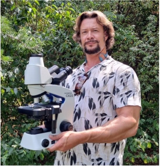 |
Q: What does your winning image show?
Igor: The image shows a morning glory pollen grain germinating on the flower's stigma. A pollen tube is emerging from one of the grain's apertures. It travels down the pistil and into the ovary in search of an egg cell. The colors are false—I rendered the image as a depth color-coded projection. The parts closer to the viewer are shown in lighter hues.
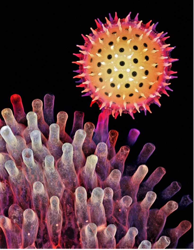
Captured by Igor Siwanowicz, this image of a morning glory pollen grain won the regional prize for the Americas in our Image of the Year 2022 competition.
Q: What do you find exciting about the image and subject matter?
Igor: I am fascinated with the intricacies of natural forms and the multiple facets of design found in nature. In summer 2022, I decided to give flowers, especially those with interestingly shaped pollen grains, some love and attention. Janelia Research Campus—where I work and live—has many species of asters and bindweeds. I didn’t need to look far to find specimens for my weekend project.
Q: How did you create the image?
Igor: I collected several stigmas (a stigma is a flower's female part; specifically, the part of a pistil where pollen germinates) from morning glory flowers growing on my institute's communal garden fences. After embedding them in 7% agarose, I sliced the specimens into 0.2 mm sections, then stained them with calcofluor-white and Congo red—cellulose-binding dyes that originate from the textile industry and have been in use (as an optical whitener and a red dye, respectively) until their carcinogenic properties were discovered. I imaged the specimen using a 40X objective on a confocal microscope.
Q: What challenges did you encounter when capturing this image?
Igor: I used similar sample prep and imaging protocols before. It was like executing a familiar recipe for a relatively simple dish. I enjoy the process of sample preparation in a similar way as cooking. In both cases, I’m mostly following my intuition and don’t sweat the details.
Q: Why did you choose this image as your entry for the Image of the Year competition?
Igor: I thought it met the criteria favored by the judges, as I learned from both browsing through past winning entries and participating in various photomicrography competitions over the past decade or so. It has a sufficiently high level of detail to make it interesting, has no blown up, overexposed, or clipped areas, follows the rules of composition, and tells a story or shows a biological phenomenon/process.
Q: Is there a message about the image that you would like to share?
Igor: There is beauty in nature on every scale, and it is ubiquitous. You can find it pretty much everywhere and sometimes in unexpected places, such as inside a snail’s mouth (a snail radula, or a rasping tongue, won me an honorable mention last year).
Of course, the beauty is in the eyes of the beholder, and one needs to approach those unfamiliar, alien-looking, and sometimes very abstract forms with an open mind. I think open-mindedness is an essential trait, or mindset, in learning and creativity. It comes with a humble confidence: the courage to admit that parts of one’s worldview could be wrong or even missing. Unfortunately, there is a tendency in some cultures to mistake arrogance and assertiveness for confidence. Refusing to give in is seen as a virtue, while changing one’s mind is perceived as a weakness.
Surely, I’m in good company when I say that I see certitude as detrimental to creativity, learning, and personal growth. The threat of certitude can only be countered by leaving the comfort and safety of knowing and cultivating a beginner’s mindset—a childlike curiosity unbiased by preconceptions.
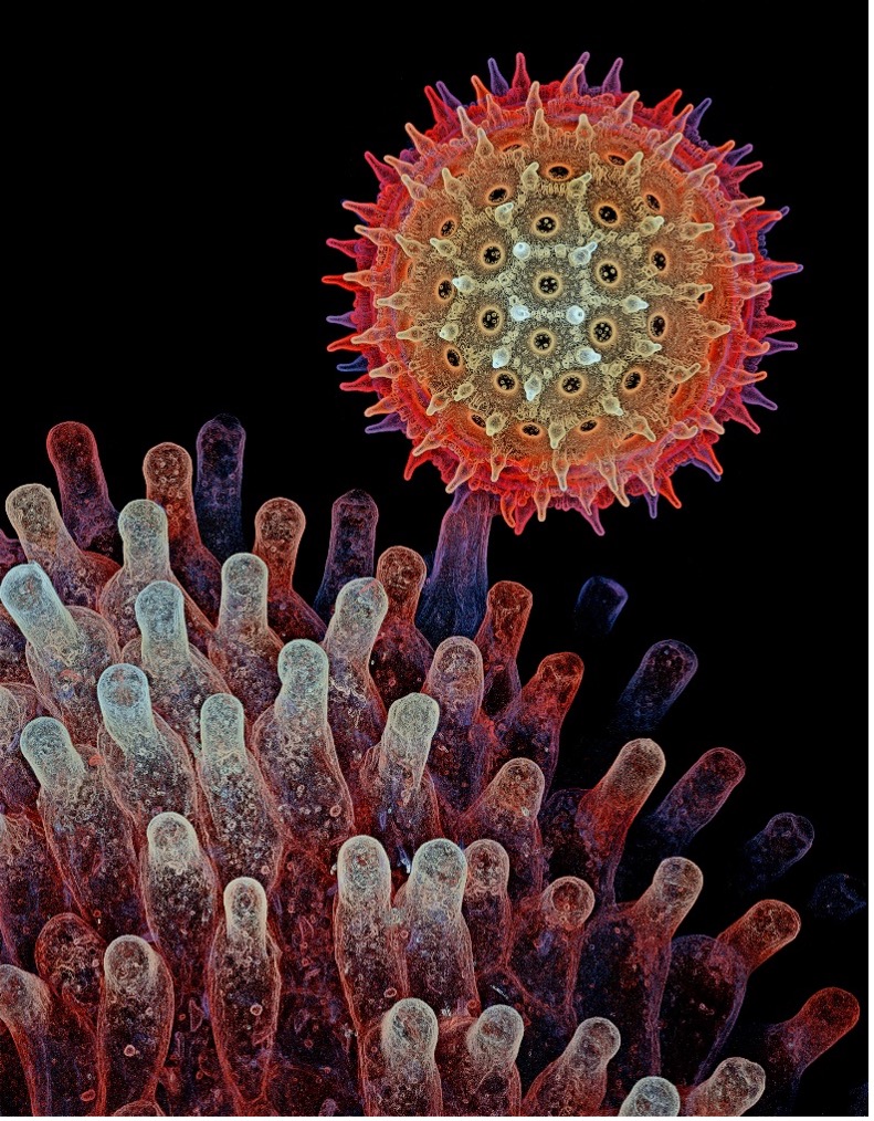
An alternative rendering of the winning image. For this depth color-coded projection, the find edges filter was applied to the stack before flattening it and creating the projection. Image courtesy of Igor Siwanowicz.
Q: Where and when did you first learn to use a microscope?
Igor: My parents had an antique brightfield microscope with a brass body and concave mirror for illumination. I was playing with it a lot as a kid, watching protozoans, rotifers, and other microfauna in water samples. But parts of it broke, and the microscope was converted into a fancy candlestick holder. It has served this function for the past 30 years or so.
I later earned a PhD in molecular biology, which involved using X-ray crystallography to solve molecular structures of proteins at the atomic level. At this magnification, while still interesting, sometimes even beautiful nature becomes abstract. Eventually, love for nature and a desire to see the bigger picture drove me to shift to the field of neurobiology. That’s when I got access to confocal microscopes.
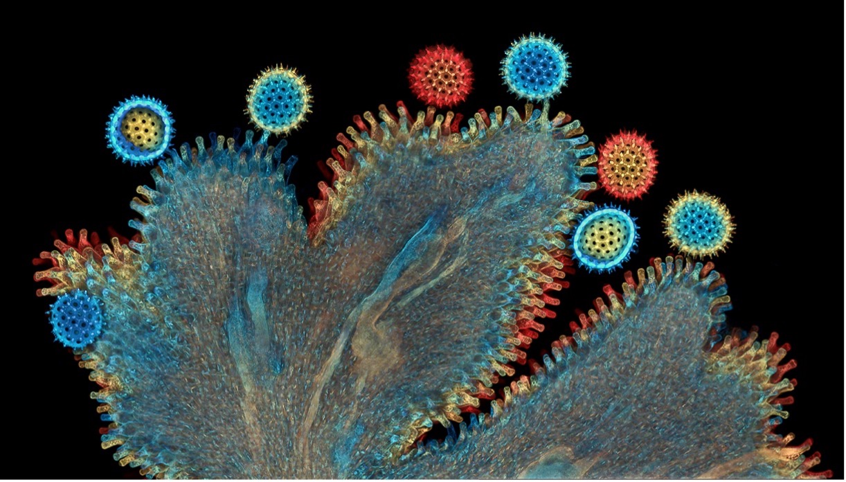
An image of the morning glory stigma at lower magnification (10X objective). Image courtesy of Igor Siwanowicz.
Q: When did you become inspired to use microscopes to create art?
Igor: My parents, both biologists, instilled in me an interest in nature and biology and primed my curiosity at an early age. I grew up surrounded by biology-related books, and I browsed through them even before I learned how to read. That’s when I first encountered the work of Ernst Haeckel. His Art Forms in Nature, a magnificent collection of highly detailed lithographs showing all sorts of life forms, is a great example of the marriage of scientific approach and artistic talent. His work remains one of my most influential inspirations.
Around 20 years ago, I bought my first camera and found myself on the supply side of nature photography, with a special focus on the macro technique. My favorite models were invertebrates, reptiles, and amphibians. I started with a defined goal: to instil in the viewers a sense of wonder, admiration, and respect for life in all its manifestations. I went about it by treating my models as miniature celebrities, meeting them eye-to-eye as an equal and applying glamour portrait photography techniques, such as soft illumination and pastel backdrops. Microscopy perfectly complemented macro photography and gave me an even more intimate perspective on natural forms.
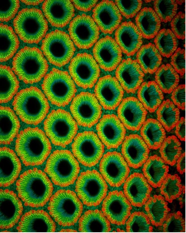
A close-up look at a damselfly eye. Image courtesy of Igor Siwanowicz.
Q: How long have you been creating art with a microscope?
Igor: While working on my PhD and during two years of postdoctoral research, I was studying interactions of proteins on the atomic level. At some point, I found it too reductionist for my taste; by focusing on a single interaction within a large biochemical pathway, one of thousands within a cell, I’m losing the bigger picture.
Around sixteen years ago, I changed my research field to one more in tune with my naturalist’s interests. Neurobiology is the study of cells in the nervous system and the organization of these cells into functional circuits that process information and mediate behavior. Insects, with a relatively simple—and hence easier to study—nervous system, are commonly used as models. On the most basic level of relatively simple neuronal networks, all nervous systems have a lot in common. It was my extracurricular expertise in invertebrate anatomy and macro photography that made the transition possible.
One of the tools often used by neurobiologists for studying the morphology of neurons and their networks is a microscope. As a bonus, I gained access to high-end microscopes, such as the confocal microscope.
Q: What do you find most fascinating about microscopy?
Igor: For most non-experts, microscopy images, with their unfamiliar and abstract shapes, may cause confusion. Hopefully this is the good kind of confusion: the kind that leads to learning. It is sometimes referred to as optimal confusion and is thought to be a key epistemic emotion, since it leads to knowledge acquisition. Confusion like that causes a certain degree of discomfort: a mental itch that comes with the recognition of a gap in one’s knowledge. That itch can only be scratched by finding the information that will close that gap.
Awe and wonder are emotions often experienced in response to things like music, art, vast landscapes, the northern lights, or a lightning storm. The size of the phenomenon doesn’t need to be a defining criterium. For me, it is the exploration of the fine details and intricacies of natural forms and designs that often trigger those emotions. Awe and wonder are both vital states of mind in our human experience. Both are humbling. They make us feel small yet connected—to ourselves, others, nature, and the universe. While awe makes us stop and take it all in, wonder inspires a desire to understand, to engage with the object or phenomenon. Wonder sparks active interest and incites deeper curiosity.
Microscopy can awaken viewers to the beauty of natural forms and the many facets of design found in nature. It has power to incite interest, spark curiosity, and motivate passionate investigations into the natural world.
Q: Does your profession intersect with imaging? Or is this more of a hobby, art form, or passion for you?
Igor: For the past 11 years, I’ve been working as a research scientist at Janelia Research Campus of the Howard Hughes Medical Institute. Among other projects, I am currently working with collaborators to develop a musculoskeletal model of the fruit fly.
I am incredibly lucky and privileged, as I was able to combine my hobby, passion, and career. Over the years, I have collaborated with labs inside and outside of Janelia to study the anatomy and neurocircuitry of invertebrates. My specialization is preparing and imaging atypical samples, in most cases containing elements of the exoskeleton, muscles, and mechanosensory or motor neurons.
Examples of my research work include:
- Controlling motor neurons of every muscle for fly proboscis reaching
- 3D reconstruction of energy stores for jumping in planthoppers and froghoppers from confocal laser scanning microscopy
- Systematic characterization of wing mechanosensors that monitor airflow and wing deformations
- Biomechanical origins of proprioceptor feature selectivity and topographic maps in the Drosophila leg
Q: What you are currently working on artistically?
Igor: My list of fun projects includes imaging sections of dragonfly eyes stained for nuclei, actin, and tubulin; Aspergillus mold and Dictyostelium slime mold raised on agar, embedded in more agar, and sectioned in a way that would show the organism from the side. I haven’t seen that done, so I’m excited about it.
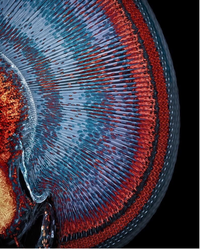
A maximum intensity projection of dragonfly eye sections. Image courtesy of Igor Siwanowicz.
Q: Is there anything else you’d like to share about your experience in the Evident Image of the Year contest?
Igor: My experience and all the interactions were very positive, of course! I am grateful Evident is bringing the wonders of the natural world to a broader audience’s attention and hope the images spark some interest and curiosity!
Related Content
Art of Chemistry: Meet the First IOTY Materials Science Award Winner
Breadth of Beauty—Our Most Popular Microscope Images for August 2023
Awards Season—Our Most Popular Microscope Images for April 2022