
Pregunte a los expertos
Nos complace recibirlo en este exclusivo portal, denominado Pregunte a los expertos, para que usted pueda hacernos sus consultas relativas al ámbito de la microscopía en las ciencias biológicas. Únase a los seminarios web en vivo dirigidos por nuestros expertos para discutir temas de vanguardia o comuníquese con ellos en cualquier momento para plantear sus preguntas y desafíos. ¿No pudo asistir a un seminario web? No se preocupe, es posible escucharlo nuevamente. ¿No se lista el tema de su interés? Utilice nuestro formulario de comentarios o póngase en contacto directo con nuestros expertos para obtener ayuda.
Seminarios web en vivo | Seminarios web por solicitud | Expertos
Accelerating Image Analysis with TruAI™ Deep Learning Technology
Expertos

Manoel Veiga
Especialista de aplicaciones - Investigación en Ciencias de la Vida
Olympus Soft Imaging Solutions
In this tech talk, through a collection of examples measured with our live cell imaging systems, high-content screening station, and whole slide scanner, you will see what TruAI technology can do for your research and get a preview of what is coming next.
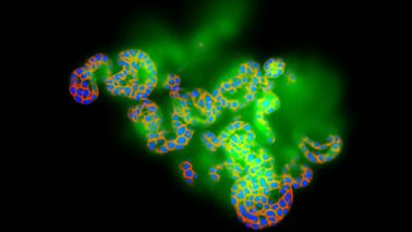
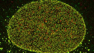
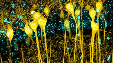
Metabolic Imaging in Langerhans Human Islets with MPE and FLIM
Expertos

Capturing life (mis)regulation at the nanoscale is a crucial challenge for present and future biophysics. At this scale, the main actors are the molecules. To successfully tackle molecular behavior within living matter, optical microscopy is a valuable methodological platform: by using fluorescence as the signal, spatial and temporal details of molecular processes can be investigated quantitatively. The physiopathology of beta-cell response to glucose stimulation will be used as case study of biological/biomedical interest. The metabolic traits of beta cells will be highlighted using a straightforward combination of multiphoton microscopy, fluorescence lifetime imaging, and feedback-based orbital tracking of sub-cellular nanostructures.
Product Demo: IXplore™ SpinSR Confocal Super Resolution System
Expertos

Stefan Marawske
Especialista sénior en ventas - Sistemas de última generación para Ciencias de la Vida
Olympus Europa
In this live demo, experience the IXplore SpinSR system, designed for fast 3D super resolution imaging and prolonged cell viability in time-lapse experiments. The microscope system offers XY resolution down to 120 nm without the need for dedicated labeling procedures. Learn how to easily integrate the IXplore SpinSR microscope system into existing experiments and sample protocols to streamline your research.
Deconvolution of 3D Image Stacks
Expertos

Images taken with a light microscope are never true representations of the specimen. Error sources, which have to be controlled, are sample preparation and staining protocols as well as optical aberrations and limitations of microscope and digital camera.

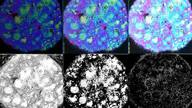
Deep Learning Approaches to Automated Phenotypic Profiling
Expertos

Quantifying cellular phenotypes is the key to all cell biology studies. However, modern imaging techniques can easily generate more data than an average user can comfortably handle. In this presentation, Dr. Chao discusses two deep learning approaches, one semi-supervised and one supervised, for building image analysis pipelines. Either approach can be run on a free cloud GPU instance.
Confocal Microscopy and Its Use for a Spaceflight Experiment
Expertos

Spaceflight experiments represent a rare but exciting scientific opportunity. Unlike most lab experiments, in which protocols can be quickly modified, limitations on crew time and availability of supplies are notable factors. Unanticipated changes to launch and reentry schedules are also an issue. The experimental apparatus and protocols used must be able to function in a microgravity setting, while also resisting the g-forces and vibrations during launch and landing. During this presentation, Dr. McLean will go over the experimental planning and use of confocal and electron microscopy approaches and analyses during a recent spaceflight experiment that flew on Space X-21 from 12/6/20 – 1/14/21.
Evolution of Scientific Digital Imaging Technologies and their Applications
Expertos

Lin Guo
Manager, Product and Application Life Science Department Scientific Solutions Business Division
Olympus Singapore
In this talk, Dr. Lin covers some critical facts about scientific digital cameras. He also discusses the evolution of these cameras, the solutions that Olympus offers, and how they are used in current advanced microscopy systems for various applications.
A New Way of Thinking—Object Detection with Deep Learning
Expertos

In this session, we will discuss object segmentation with deep learning and its applications in life science. We will also demo Olympus deep-learning software.
Product Demo: SLIDEVIEW™ VS200 Research Slide Scanner
Expertos

In this product demo you will learn how to capture high-resolution images of your slides for quantitative analysis, enabling you to make the most of the information your slides have to offer. Easily analyze, share, and archive your data with the SLIDEVIEW VS200 digital slide scanner. Join this session and learn how to achieve more in less lead time.
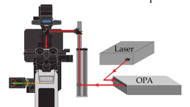
In-Vivo Tracking of Harmonic Nanoparticles by Means of a TIGER Widefield Microscope
Expertos

In-vivo tracking based on harmonic nanoparticles is so far not exploited because of a lack of an appropriate tool—a widefield nonlinear optical microscope. Here, we present a new approach to overcome this challenge based on a redesign of laser space parameters.
Product Demo: FLUOVIEW™ FV3000 Confocal Laser Scanning Microscope
Expertos

Join Bülent Peker, Senior Product Marketing Manager to see how the FV3000 confocal laser microscope can expand your research possibilities and help you get more data from your samples.
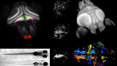
Whole-Brain Functional Calcium Imaging Using Light Sheet Microscopy
Expertos

Light sheet microscopy is a powerful technique to perform fast volumetric imaging. I will talk about how we use it to investigate how the brain of a small vertebrate, the larval zebrafish, works. In our laboratory at the Technical University of Munich, we are interested in how the brain processes external sensory stimuli and uses internal states and past experiences to select appropriate behavior. In order to do this, we image the activity of almost all 100,000 neurons in the brain of larval zebrafish while we present the animals with stimuli and monitor their behavior. I will also discuss the data processing steps after acquiring these large datasets.