The substage condenser gathers light from the microscope light source and concentrates it into a cone of light that illuminates the specimen with uniform intensity over the entire viewfield. It is critical that the condenser light cone be properly adjusted to optimize the intensity and angle of light entering the objective front lens. Each time an objective is changed, a corresponding adjustment must be performed on the substage condenser to provide the proper light cone for the numerical aperture of the new objective.
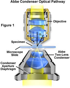
A simple two-lens Abbe condenser is illustrated in Figure 1. In this figure, light from the microscope illumination source passes through the condenser aperture diaphragm, located at the base of the condenser, and is concentrated by internal lens elements, which then project light through the specimen in parallel bundles from every azimuth. The size and numerical aperture of the light cone is determined by adjustment of the aperture diaphragm. After passing through the specimen (on the microscope slide), the light diverges into an inverted cone with the proper angle to fill the front lens of the objective.
Condenser Aperture Diaphragm Operation
Explore how the substage condenser aperture controls illumination entering the objective.
Aperture adjustment and proper focusing of the condenser are of critical importance in realizing the full potential of the objective. Specifically, appropriate use of the adjustable aperture iris diaphragm (incorporated into the condenser or just below it) is most important in securing correct illumination, contrast, and depth of field. The opening and closing of this iris diaphragm controls the angle of illuminating rays (and thus the aperture) which pass through the condenser, through the specimen and then into the objective. Visitors are invited to explore how changing the condenser aperture effects the illumination cone in our interactive Java tutorial that explores the condenser numerical aperture. Condenser height is controlled by a rack and pinion gear system that allows the condenser focus to be adjusted for proper illumination of the specimen. Correct positioning of the condenser with relation to the cone of illumination and focus is critical to quantitative microscopy and optimum photomicrography.
Care must be taken to guarantee that the condenser aperture is opened to the correct position with respect to objective numerical aperture. When the condenser aperture diaphragm is opened too wide, stray light generated by refraction of oblique light rays from the specimen can cause glare and lower the overall contrast. On the other hand, when the aperture is made too small, the illumination cone is insufficient to provide adequate resolution and the image is distorted due to refraction and diffraction from the specimen. Visitors can explore these phenomena with our Interactive Condenser Aperture Java Tutorial that demonstrates the effect of condenser aperture position on specimen illumination.
Condenser Effects on Image Contrast
Discover how the size of the condenser aperture affects specimen image contrast.
Condensers are divided into classifications of purpose (e.g.: brightfield, darkfield, phase contrast, etc.), and also according to their degree of optical correction. There are four principle types of condensers with respect to correction of optical aberrations, as listed in Table 1.
Condenser Aberration Corrections
| Condenser Type | Aberrations Corrected | |
|---|---|---|
| Spherical | Chromatic | |
| Abbe | --- | --- |
| Aplanatic | x | --- |
| Achromatic | --- | x |
| Aplanatic- achromatic | x | x |
Table 1
The simplest and least corrected (also the least expensive) condenser is the Abbe condenser that can have a numerical aperture up to 1.4 in high-end models with three or more internal lens elements. While the Abbe condenser is capable of passing bright light, it is not corrected for either chromatic or spherical optical aberrations. A typical Abbe condenser is illustrated in Figure 2. In its simplest form, the Abbe condenser has two optical lens elements that produce an image of the illuminated field diaphragm that is not sharp and is surrounded by blue and red color at the edges.
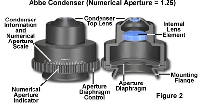
As a result of no optical correction, the Abbe condenser is suited mainly for routine observation with objectives of modest numerical aperture and magnification. The primary advantages of the Abbe condenser are the wide cone of illumination that the condenser is capable of producing as well as its ability to work with long working distance objectives. Most microscopes are supplied by the manufacturer with an Abbe condenser as the default and these condensers are real workhorses for routine laboratory use.
The next level of condenser correction is split between the aplanatic and achromatic condensers that are corrected exclusively for either spherical (aplanatic) or chromatic (achromatic) optical aberrations. Typical examples of these condensers are illustrated in Figures 3 (achromatic) and Figure 4 (aplanatic). Achromatic condensers usually contain three to four lens elements and are corrected in two wavelengths (red and blue) for chromatic aberration.
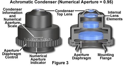
The achromatic condenser illustrated in Figure 3 contains four lens elements and has a numerical aperture of 0.95, the highest attainable without requiring immersion oil. This condenser is useful for both routine and critical laboratory analysis with "dry" objectives and also for black and white or color photomicrography.
A critical factor in choosing substage condensers is the numerical aperture performance that will be necessary to provide an illumination cone adequate for the objectives. The condenser numerical aperture should be equal to or slightly less than that of the highest objective numerical aperture. Therefore, if the highest magnification objective is an oil-immersion objective with a numerical aperture of 1.40, then the substage condenser should also have an equivalent numerical aperture to maintain the highest system resolution. In this case, immersion oil would have to be applied between the condenser top lens and the underside of the microscope slide to achieve the intended numerical aperture (1.40) and resolution. Failure to use oil will restrict the highest numerical aperture of the system to 1.0, the highest obtainable with air as the imaging medium.
Transmitted Microscopy Light Pathways
Discover how the condenser and field diaphragms affect illumination in transmitted microscopy.
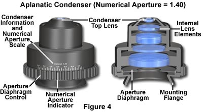
Aplanatic condensers are well corrected for spherical aberration (green wavelengths) but not for chromatic aberration. A typical aplanatic condenser with a numerical aperture of 1.40 is illustrated in Figure 4. This condenser features five lens elements and is capable of focusing light in a single plane. Aplanatic condensers are capable of producing excellent black and white photomicrographs when used with green light generated by either a laser source or by use of an interference filter with tungsten-halogen illumination.
The highest level of correction for optical aberration is incorporated in the aplanatic-achromatic condenser. This condenser is well corrected for both chromatic and spherical aberrations and is the condenser of choice for use in critical color photomicrography with white light. A typical aplanatic-achromatic condenser is illustrated in Figure 5 (numerical aperture = 1.35). This condenser features eight internal lens elements cemented into two doublets and four single lenses.

Engravings found on the condenser housing include its type (achromatic, aplanatic, etc.), the numerical aperture, and a graded scale that indicates the approximate adjustment (size) of the aperture diaphragm. As we mentioned above, condensers with numerical apertures above 0.95 perform best when a drop of oil is applied to their upper lens in contact with the undersurface of the specimen slide. This ensures that oblique light rays emanating from the condenser are not reflected from underneath the slide, but are directed into the specimen. In practice, this can become tedious and is not commonly done in routine microscopy, but is essential when working at high resolutions and for accurate photomicrography using high-power (and numerical aperture) objectives.
Another important consideration is the thickness of the microscope slide, which is as crucial to the condenser as coverslip thickness is to the objective. Most commercial producers offer slides that range in thickness between 0.95 and 1.20 mm with the most common being very close to 1.0 mm. A microscope slide of thickness 1.20 mm is too thick to be used with most high numerical aperture condensers that tend to have a very short working distance. While this does not greatly matter for routine specimen observation, the results can be devastating with precision photomicrography. We recommend that microscope slides be chosen that have a thickness of 1.0 ± 0.05 mm, and that they be thoroughly cleaned prior to use.
Condenser Light Cones
Study how optical correction affects the size and shape of condenser light cones.
When the objective is changed, for example from a 10X to 20X, the aperture diaphragm of the condenser must also be adjusted to provide a new light cone that matches the numerical aperture of the new objective. This is done by turning the knurled knob on the condensers illustrated in Figures 2-6. There is a small yellow arrow or index mark located on this knob that indicates the relative size of the aperture when compared to the linear gradation on the condenser housing. Many manufacturers will synchronize this gradation to correspond to the approximate numerical aperture of the condenser. For example, if the microscopist has selected a 10X objective of numerical aperture 0.25, then the arrow would be placed next the value 0.18-0.20 (about 80 percent of the objective numerical aperture) on the gradation inscribed on the condenser housing.
Often, it is not practical to use a single condenser with an entire range of objectives (2X to 100X) due to the broad range of light cones that must be produced to match objective numerical apertures. With low-power objectives in the range 2X to 5X, the illumination cone will have a diameter between 6-10 mm, while the high-power objectives (60X to 100X) need a highly focused light cone only about 0.2-0.4 mm in diameter. With a fixed focal length, it is difficult to achieve this wide range of illumination cones with a single condenser.
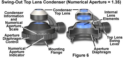
In practice, this problem can be solved in several ways. For low power objectives (below 10x), it may be necessary to unscrew the top lens of the condenser in order to fill the field of view with light. Some condensers are produced with a flip-top upper lens to accomplish this more readily, as illustrated in Figure 6. Many manufacturers now produce a condenser which flips over completely when used with low power objectives. Other companies may incorporate auxiliary correction lenses in the light path for securing proper illumination with objectives less than 10x. When the condenser is used without its top lens, the aperture iris diaphragm is opened wide and the field diaphragm, now visible at the back of the objective, serves as if it were the aperture diaphragm. Flip-top condensers are manufactured in a variety of configurations with numerical apertures ranging from 0.65 to 1.35. Those condensers that have a numerical aperture value of 0.95 or less are intended for use with "dry" objectives. However, flip-top condensers that have a numerical aperture greater than 0.95 are intended for use with oil-immersion objectives and they must have a drop of oil placed between the bottom of the microscope slide and the condenser top lens when examining critical samples.
In addition to the common brightfield condensers discussed above, there are a wide variety of specialized models suited to many different applications. Table 2 lists a number of different condenser configurations and the intended applications.
Substage Condenser Applications
| CONDENSER TYPE | BRIGHTFIELD | DARKFIELD | PHASE CONTRAST | DIC | POLARIZING |
| Achromat/ Aplanat N.A. 1.3 | ⋅ [10x~100x] | ||||
| Achromat Swing-out N.A. 0.90 | ⋅ [4x~100x] | ||||
| Low-Power N.A. 0.20 | ⋅ [1x~10x] | ||||
| Phase Contrast Abbe N.A. 1.25 | ⋅ | ⋅ [up to N.A. 0.65] | ⋅ [10x~100x] | ||
| Phase Contrast Achromat N.A. 0.85 | ⋅ | ⋅ [up to N.A. 0.70] | ⋅ [4x~100x] | ||
| DIC Universal Achromat/Aplanat | ⋅ | ⋅ [up to N.A. 0.70] | ⋅ [10x, 100x] | ⋅ [20x, 40x, 100x] | |
| Darkfield, dry N.A. 0.80~0.95 | ⋅ [4x~40x] | ||||
| Darkfield, oil N.A. 1.20~1.43 | ⋅ [4x~100x] | ||||
| Stain-Free Achromat Swing-Out N.A. 0.90 | ⋅ | ⋅ [4x~100x] |
Table 2
From the data in Table 2, it is obvious that substage condensers have a great deal of interchangeability among different applications. For instance, the DIC universal achromat/aplanat condenser is useful for brightfield, darkfield, and phase contrast, in addition to the primary DIC application. Other condensers have similar interchangeability. We will deal with various aspects of individual techniques that require modified condensers in our section on Specialized Microscopy Techniques. Please feel free to visit this interesting part of our Website for more specific information about substage condensers.


