Guidance for Quantitative Confocal Microscopy
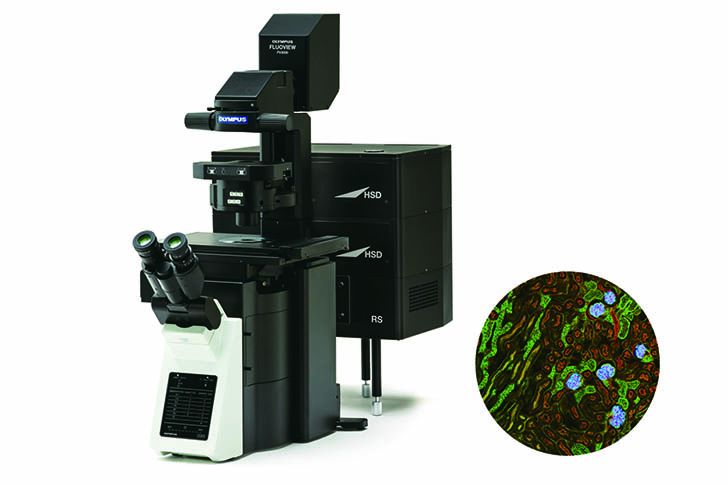
When used correctly, a confocal fluorescence microscope is an excellent tool for making quantitative measurements in cells and tissues. Its ability to block out-of-focus light and thereby perform optical sectioning through a specimen enables you to quantify fluorescence with very high spatial precision.
However, generating meaningful data using confocal microscopy requires careful planning and a thorough understanding of the technique. This webinar will guide you through key aspects of acquiring quantitative confocal microscopy images, including:
- Optimizing sample preparation
- Choosing the most suitable microscope for a given application
- Configuring the microscope parameters
You’ll also learn about common pitfalls such as photobleaching and crosstalk, as well as several instrument problems that may prevent the acquisition of quantitative data.
The webinar is based on a recent Nature Protocols tutorial paper authored by an international group of core microscopy facility directors: James Jonkman, Claire M. Brown, Graham D. Wright, Kurt I. Anderson, and Alison J. North. Tutorial: Guidance for Quantitative Confocal Microscopy. Nature Protocols. (2020) 15: 1585-1611.
Presenters:
 | Dr. Graham Wright, Chief Technology Officer |
Guidance for Quantitative Confocal Microscopy
|
このページはお住まいの地域ではご覧いただくことはできません。