소프트웨어
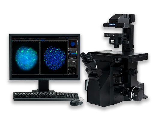
Discover more with Olympus microscope software. With intuitive operations that offer a seamless workflow, Olympus microscope software supports your ever-evolving research needs. Digital imaging microscope software provides a vast range of features for different applications, whether you’re looking for simple snapshot images or are performing advanced multi-dimensional, real-time experiments. Olympus microscope software enables image acquisition in five dimensions, along with powerful analysis tools that dynamically extract data from imagery for precise experiment results. Confluency checker microscope software is also a must for users looking to improve the quality of cell culture processes with quantitative analysis. Confluency checker software is used alongside images captured with your digital microscope to automatically count both the number of cells and the percent of confluency in culture vessels. Olympus confluency microscope software offers fast, precise cell counting, providing you with quantitative growth data to improve your cell culture process. |
|
Microscope Software
Maximum Compare Limit of 5 Items
Please adjust your selection to be no more than 5 items to compare at once
Microscope Software Resource Videos
cellSens Software Deep Learning Part 1. Preparing Training DataRather than using fluorescence images to detect and count nuclei, deep-learning technology can count the nuclei using simple transmitted images, with no staining. This cellSens tutorial video shows you how to use cellSens software’s deep-learning technology for label-free nucleus detection. | 관련 영상 |
Macro to Micro ImagingDiscover how macro to micro imaging can make your experiments more efficient and reliable. This video explores how A.I. technology uses neural networks to automatically detect your samples when traditional contrast methods fall short. | 관련 영상 |
Not Available in Your Country
Sorry, this page is not
available in your country.