
New year, new images! Our top images from January highlight a mix of traditional research samples and nontraditional imaging techniques. Capturing cells to crystalized chemicals, these images boast colors across the full spectrum. See all the favorites below.
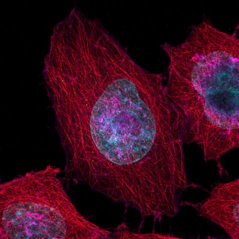
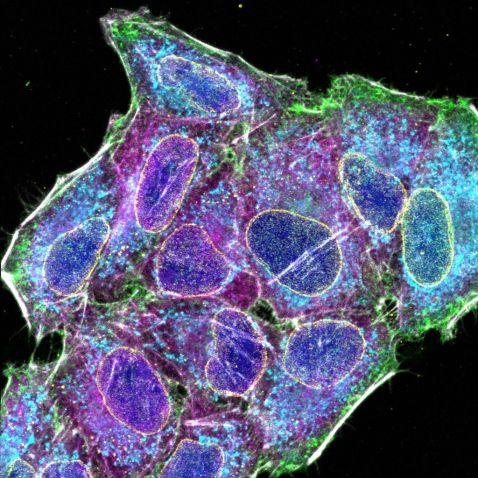
Multiplexing, the process of combining two or more information channels into a single transmission medium, has grown more popular and more important in recent years. By imaging only a small sample, researchers can view details of varied targets and interest points in parallel. Our most popular image this month demonstrates this technique. HeLa* cells from a cytoskeleton sample were stained with DAPI (blue), Pericentrin (centrosome, green), a-tubulin (microtubules, Alexa 568; red), and phalloidin (actin, Alexa 647; magenta).
Sample preparation courtesy of Alexia Ferrand. Sample acquisition courtesy of Sara R. Roig and Alexia Ferrand, Imaging Core Facility, Biozentrum, University of Basel. Captured using a FLUOVIEW™ FV4000 confocal laser scanning microscope.
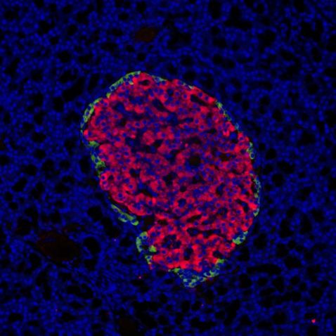
Are we looking at a post-impressionist painting? While this image reminds us of the Pointillism style of painting pioneered by Georges Seurat and Paul Signac, it’s actually an image of a rat pancreas stained with DAPI AF555 Cy5. Located in the abdomen, the pancreas is an organ that aids in endocrine and digestive functions.
Captured using a BX53 microscope with a DP75 camera.
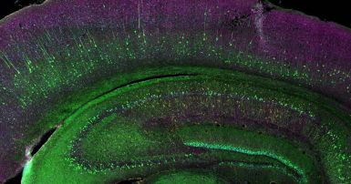
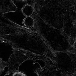
The human brain is vastly larger than that of a mouse, but the types of neurons and connections within them are quite similar. This makes them valuable research subjects. In addition to being scientifically important, mouse brain slices make for stunning images.
Here we can see the coronal section of an H-line mouse brain. The sample was stained with cyan; DAPI (cell nuclei), green; YFP (neuron), yellow; Cy3 (astrocytes), magenta; and Alexa Fluor 750 (microtubule).
Sample courtesy of Takako Kogure and Atsushi Miyawaki, Cell Function Dynamics, RIKEN CBS. Captured using an FV4000 confocal laser scanning microscope.

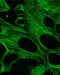
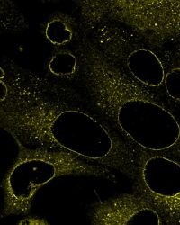
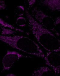
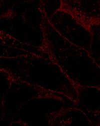
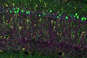
Here we see another series of images of HeLa cells, in this case labeled by six fluorochromes: cell nuclei (DAPI; blue), cell membrane (AF 488; green), nuclear pore (AF 561; yellow), microtubule (Qdot 605; magenta), mitochondria (MitoTracker DeepRed; cyan), and actin (AF 750 phalloidin; gray). This provides us with a beautiful rainbow of colors and a broad view of the various aspects of the sample.
And as a bonus, here’s the multiplexed version!
Images captured using an FV4000 confocal laser scanning microscope.
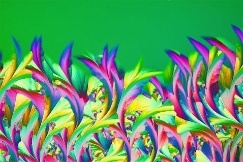
Are we in a modern art museum? This colorful image also reminds us of a painting.
“Consider the image above. At first glance, it may look like an abstract painting. However, it actually shows the crystal of a topical medicine for wart treatment, also known as callus remover. You can see the pretty pattern that evolved during the crystallization process of the substance on the microscope slide.”
To create this image, Shyam Rathod put a drop of callus remover, which is a combination of salicylic acid and lactic acid, on a slide. The drop was then pressed with another slide and gently moved to form a thin layer. A retarder and a two-cross polarization filter were used to bring out the colors, which were captured in a single frame. To learn more about crystal art photomicrography, read Shyam’s guest post discussing the process.
Image and quote courtesy of Shyam Rathod, materials science winner of the Evident Image of the Year 2022 competition. To learn more about Shyam’s winning image, read our interview. To see all the winning images, visit olympus-lifescience.com/ioty.
To see more images like these, be sure to follow us on Instagram at @evidentlifescience!
Interested in sharing your own images? Visit our image submission site!
*Although it became one of the most important cell lines in medical research, it’s imperative that we recognize Henrietta Lacks’ contribution to science happened without her consent. This injustice, while leading to key discoveries in immunology, infectious disease, and cancer, also raised important conversations about privacy, ethics, and consent in medicine. To learn more about the life of Henrietta Lacks and her contribution to modern medicine, visit the Henrietta Lacks Foundation.
Related Content
Stars and Smiles—Our Most Popular Microscope Images for December 2023
Over the Rainbow—Our Most Popular Microscope Images for November 2023
Pretty Pollen—Our Most Popular Microscope Images for October 2023
