
From bees to the pollen they love, this month’s Olympus Life Science Instagram page was buzzing with the sights of summer! Also making the top 5 this month is a response to our Sci Art Challenge.
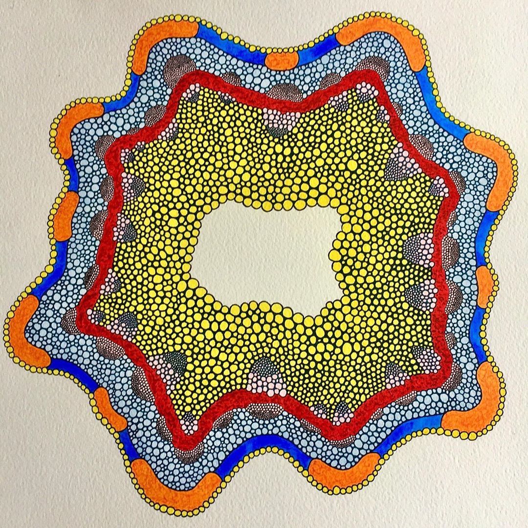
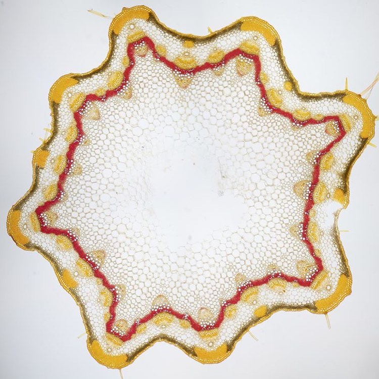
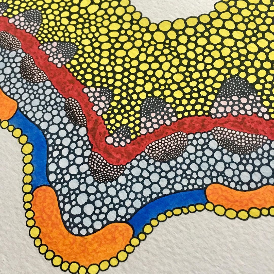
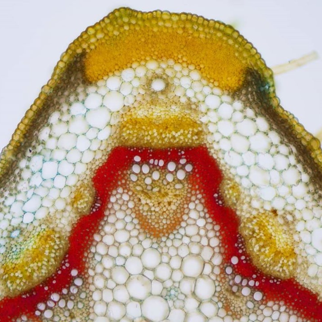
We recently issued a Sci Art Challenge to recreate our microscope images using different techniques. Our top image this week is a painting of a cross-section of a cow parsley stem originally photographed by Karl Gaff. Julia, a self-described natural sciences graduate and art enthusiast, transformed this microscope image into a work of art using gouache, watercolor, and ink to recreate it in her own style. In Karl’s original image, sections of the plant were transferred from one dye and into another with washing in between to clear color clouds.
Share your own artistic renditions of our microscope images by tagging Olympus Life Science with the hashtag #SciArtChallenge!
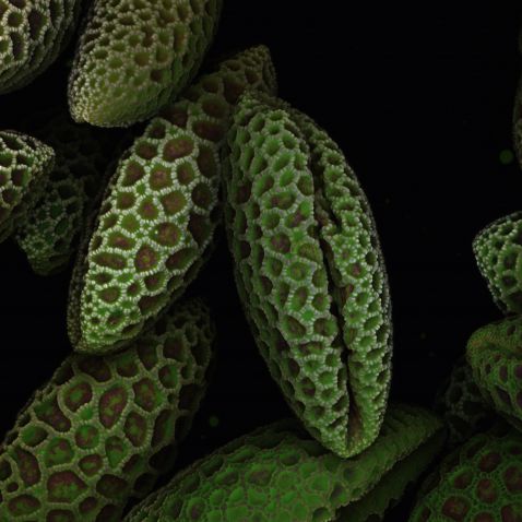
This close-up of lily pollen shows detailed textures not apparent to the naked eye when looking at the powdery (and often messy) substance.
Image captured at 40x on a FLUOVIEW FV3000 microscope by James Lopez, Manager, Olympus Life Science Applications Team.
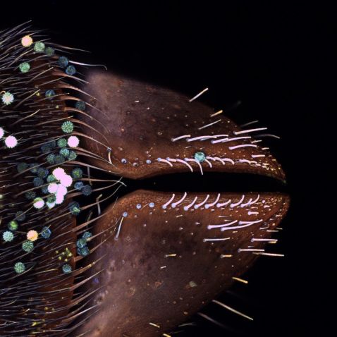
Here's a close-up look at the tube-like mouth of a bee, known as the proboscis. The proboscis has many supporting mouthparts including a pair of strong mandibles, a labrum, and two maxillae, which are lip-like structures that support the proboscis.
Image captured on a FLUOVIEW FV3000 microscope with a 10X X Line objective by Sarah Thomas, Olympus Confocal and Multiphoton Specialist.
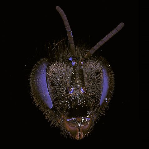
Our next most popular image takes a step back from the proboscis and captures the bee’s entire head. Fun fact: Bees can fly about 20 mph and have 2 pairs of wings to support them during flight!
Image captured on a FLUOVIEW FV3000 microscope with a 4X X Line objective by Sarah Thomas, Olympus Confocal and Multiphoton Specialist.
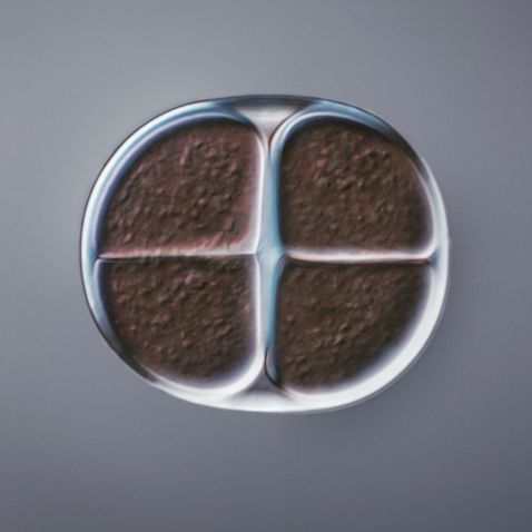
“This Chroococcus sp. (color berry) is a cyanobacteria that is widely distributed in freshwater, less in saline waters. It is found in damp places such as moist soil, tree trunks, moist walls, and rocks. The cells are single or united into spherical colonies, each containing a small number of cells surrounded by mucilage. Many species are routinely incorrectly identified, and the ecology is important for correct identification.”
Caption and image courtesy of Håkan Kvarnström. Captured on an Olympus BX51 microscope with a 60X X Line oil objective and differential interference contrast.
To see more images like these, be sure to follow us on Instagram at @olympuslifescience!
Interested in sharing your own images?
Visit our image submission site.
Related Content
Barrier-Breaking and Now Edison Award-Winning Objectives
