Not Available in Your Country
Sorry, this page is not
available in your country.
Visão geral
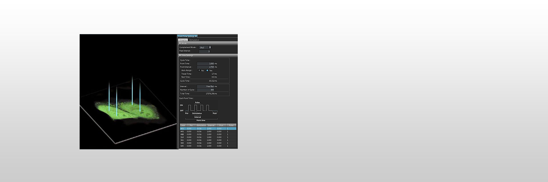
 | The Multi Point and Mapping Software Module for the FV3000 and FVMPE-RS microscopes enables the user to perform precise light stimulation of multiple points, to extend multiple points to cover a small area, or to stimulate of a rectangular area of interest for mapping scans. Stimulation timing, duration and intervals can be defined very precisely, and users can program continuous or pulse stimulation. Software control ensures that neighboring points are not excited, while synchronization and stimulation with imaging to microsecond precision allow detailed analysis of the connectivity of cells. |
|---|
Multi Point ScansThe user can designate the number of points on an image for light stimulation. Stimulation timing, duration and interval can be freely defined, and the user can program the experiment with continuous or pulse stimulation. The software also provides features that enable the stimulation of extended multiple points surrounding one single point to cover a small area. |
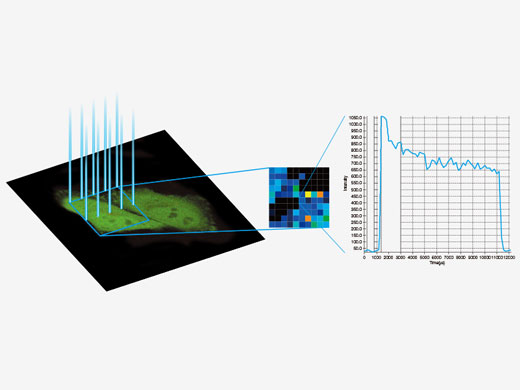
| Mapping ScansLight stimulation can be applied to a rectangular region of interest. Software control ensures that neighboring points will not be excited, enabling the user to observe the reaction of the target sample more accurately. Changes in intensity from those points can be processed as a mapped image or graph. Multiple points can be automatically set to pixels exceeding a defined threshold, helping to define a multi point scan of the active regions. These features can be combined with the IO inter-face Box for synchronization with external devices. |
|---|
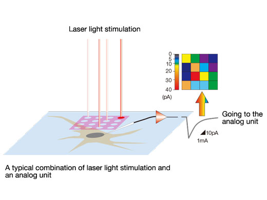
Create Activity MapsIO inter-face Box of the FV3000 converts voltages into images that can be handled just like fluorescence images. For example, electrical signals measured by patch clamping during a mapping scan can be displayed with pseudo-color and overlaid with a fluorescent image. |
|
Need assistance? |

