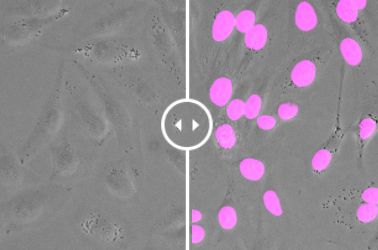
Image analysis is widely used in life science to quantify and understand events in biological samples. Object detection and segmentation are key processes for image analysis to identify our area of interest in the images. Then we can quantify morphological information, intensities, velocities in tracking, etc.
Conventional segmentation has not always been accurate and efficient; however, our eyes and brains can identify where our areas of interest are from experience. Using deep learning, we can train a neural network with ground truth information to carry out this complex task. Once the neural network has been created properly, it can help segment objects in a similar way as your brain. Deep learning sounds like it requires programming skills, but our software does not require programming skills and is easy to use.
In this session, we will discuss object segmentation with deep learning and its applications in life science. We will also demo Olympus deep-learning software.
Presenter: Akira Saito
Assistant Manager, Marketing and Applications, Olympus Singapore
Akira studied veterinary medicine at Tokyo University of Agriculture and Technology, Japan and graduated in 2007. Shortly after, he joined Olympus as application specialist responsible for in vivo imaging systems, high-content analysis systems, and laser confocal systems to support customers in Japan. In 2013, he took over sales promotion for all Olympus life science products. From 2018, he moved to Singapore and joined to support the marketing and application support for the APAC market.