Olympus provides high-quality optical darkfield components, such as the UPlanFL 60X and 100X oil iris objectives. Combined with an oil-coupled darkfield condenser, these objectives create images with improved particle detection capabilities and increased signal-to-noise ratio (SNR) when compared to standard transmitted light microscopy. Yet, the rapidly expanding needs of nanoscale research imaging have driven demand for even better darkfield performance.
An Introduction to the Enhanced Darkfield Illumination Technique
To answer this need, CytoViva introduced the enhanced darkfield (EDF) illumination technique in 2005—US patent Nos. 7,542,203, 7,564,623 (2009). This technique improves SNR performance by as much as 10X compared to standard darkfield imaging techniques. As a result, it vastly improves the detection capabilities for nanoscale entities.
CytoViva EDF illumination enables you to quickly and easily observe nanoscale entities in a range of transparent and translucent sample environments in their native state—without using labels or other markers. This capability is illustrated in Figure 1 below. The images show macrophage cells exposed to low-density lipids (LDL) loaded with gold nanoparticles (AuNPs).
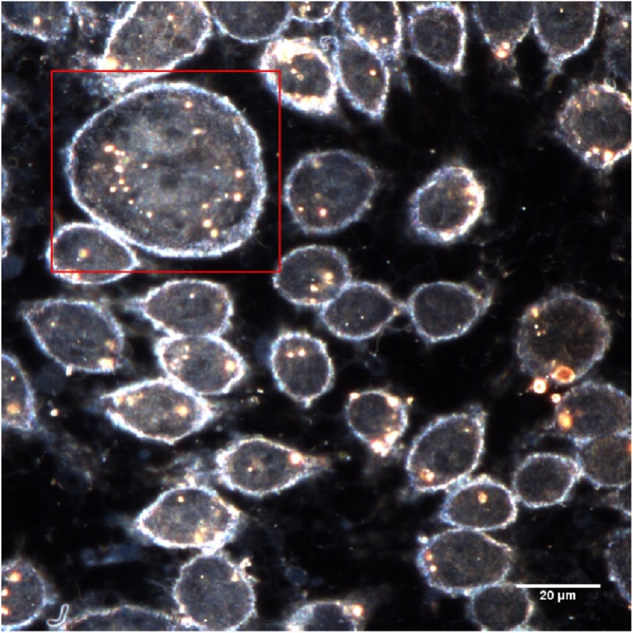
Figure 1a: Enhanced darkfield Image (60X) of LDL AuNPs in macrophage cells
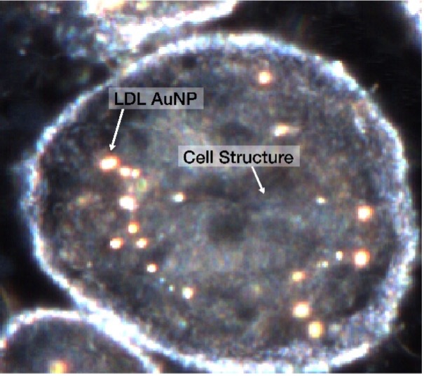
Figure 1b: 4x digital zoom of inset area highlighted in red
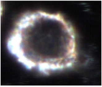
Figure 1c: Control cell
Enhanced Darkfield Illumination Opens New Possibilities in Nanoscale Imaging
This improved performance has opened new possibilities. It enables researchers to optically image nano-constructs in a variety of translucent sample matrices with enhanced clarity and fidelity. This includes:
- Plasmonic particles (Au, Ag, Pt) down to 10–20 nm
- Metal oxides (TiO2, Fe2O3, ZnO2) down to 20–40 nm
- Polymeric particles down to 40–60 nm
- Lipids down to 80–100 nm
This technique can also be used to easily image other nanoscale constructs, such as single and multi-walled carbon nanotubes. Again, no labeling or other special sample preparation is needed before imaging. Further, the technique is easy to master. New users can learn to obtain high-quality images in less than one hour, even without prior experience in microscopy.
Enhanced Darkfield Illumination: How the Technique Works
The improved performance is the result of bypassing the normal light path through the microscope base. Instead, the enclosed system and light path directs the illumination directly into the condenser, fixing the light path’s geometry from the light source to the condenser’s entrance slit (Figure 2a). This focuses the maximum photon density on the sample, enabling enhanced SNR imaging performance. Further, this design makes focusing the condenser onto the desired imaging plane of the sample easier and more consistent.
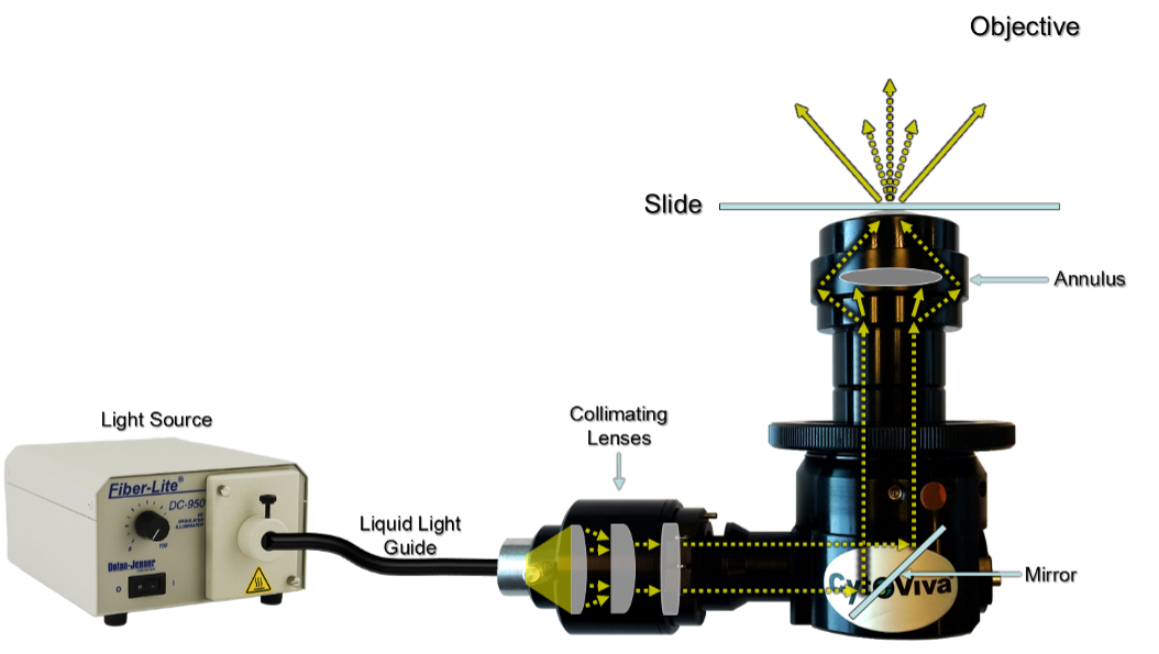
Figure 2a: Light path through the CytoViva enhanced darkfield illuminator
The EDF design also collimates the light entering the condenser. This reduces stray light and background noise. The result is an optimized darkfield image with enhanced SNR performance when compared to standard, commercially available darkfield condensers. The EDF illuminator is compatible with various Olympus upright and inverted microscopes, including:
The EDF illuminator developed by CytoViva also works with various light sources that have outputs ranging from 400–2200 nm. This includes light sources optimized for fluorescence imaging.
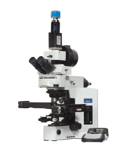
Figure 2b: CytoViva enhanced darkfield illuminator installed on an Olympus upright microscope
The enhanced darkfield illumination technique can be used across a wide variety of nano-related applications—from alternative energy to cosmetics. All these applications have their own unique requirements. Yet, they all share the need to optically observe the forming, functionalization, and location of nano-constructs in their native, non-labeled state as they are placed in various biological and non-biological matrices. As shown here, the CytoViva enhanced darkfield illuminator coupled with high-quality Olympus microscopy equipment is the key to success in these challenging applications.