
The submission period for our 2022 global Image of the Year (IOTY) contest is well underway. During this time, we have an annual tradition of sharing the stories behind the previous year’s winning images. We ask each regional winner and the global winner a series of questions to delve into their backgrounds, interest in microscopy imaging, as well as their artistic inspiration. As you’ll learn in this profile of the 2021 IOTY winner for the Asia-Pacific region, Daniel Han, microphotography is a hobby that he started surprisingly recently. Q: Where and when did you first learn to use a microscope? Do you have a scientific background?Daniel: I first used a microscope when I was working as a research assistant, but it was not until the pandemic that I actually learned how to use the instrument after purchasing a decommissioned microscope through a classified ad on a website. Before that, I built a custom measuring microscope setup to look at common objects and I also did a lot of photomacrography with it. I have a degree in electrical engineering and mathematics, so I do come from a scientific background. |  Daniel Han with his IOTY Award |
|---|
Q: When did you become inspired to use microscopes to create art?
Daniel: I started several years ago with an industrial macro lens taken from a line scanner camera that I found in an electronics bin. I wanted to get closer, so I purchased metallurgical objectives and learned how to build my own measuring microscope system, which was very good at taking microscopic photographs as well. That made me study lighting and ways to improve the images through z-stacking.
To get even closer, I chose to purchase a microscope. With my previously obtained knowledge, I automated the microscope with a stepper motor and connected it to my 35 mm format consumer camera via sets of tubing. A microscope is different, I had to study its adjustment and alignment procedures to get optimal resolution and contrast.
Q: What do you find most fascinating about microscopy, and where does this fascination stem from?
Daniel: The number of methods to generate contrast and increase resolution, and the theory behind it all is fascinating. I find it incredible that people such as Eric Betzig and his team can come up with highly sophisticated microscopy techniques such as light sheet (selective plane illumination). It was also surprising to learn that highly specialized techniques are able to break Abbe’s diffraction limit (super resolution methods such as STORM and PALM), which I was taught to be impossible in physics lessons. It was a revelation synonymous to my first encounter with imaginary numbers: “Wait! My teacher said you cannot square root a negative number!”
I suppose, being a major in mathematics taught me to appreciate the numbers and the results. All the theory behind these techniques is just fascinating to me. I also grew up fiddling with building toys, from Lego bricks to sand on a beach. This taught me to appreciate the practical side of engineering. I see harmony in many optomechanical microscopy setups.
Q: Can you tell us about your winning image and what you find most fascinating about the subject?
Daniel: The image demonstrates the back of freshly picked fern leaves, where the spore capsules (sporangium) implode, dispersing spores everywhere. Fern leaves, the sporangium, and the sorus, which is made up of numerous sporangia, auto-fluoresces when being subjected to UV light.
The most fascinating aspect of the subject was that I was able to witness the imploding spore capsules in real time under the eyepieces. After a moment of examination, the coverslip was covered with bright fluorescing fern spores. This was one of the three photomicrographs I chose to enter because it was colorful and juxtaposed intact spore capsules with an imploded one.
Q: Is there a message about the image or subject matter that you would like to communicate?
Daniel: Many common subjects will auto fluoresce. Give it a try under the microscope!
Q: Does your profession intersect with imaging or is this more of a hobby, art form, or passion for you?
Daniel: I sell and support scientific instruments and scientific imaging devices is my specialty at the company I work for. On the surface, they do intersect; however, I rarely get any hands-on experience with the sophisticated and expensive devices I deal with professionally. I must resort to the second-hand market and putting my knowledge in electronics to work for a repair. I would say it is more of a hobby and a passion for me. I love creating these photomicrographs.
I do not have any work to share as a professional, but I do have some humorous blog entries about imaging, and I have an Instagram account that is dedicated to diatoms. I also have many blog entries pertaining to the automation of my microscope.
I really love and appreciate the backwards-forwards compatibility offered by Olympus products. It is amazing to see BH2 era condensers working on a modern BX microscope, and various sliders being compatible with minimum amounts of tinkering. This just does not apply to many other brands. I have used a BX53 microscope, and now I am using an AX70 microscope. I have an IX73 microscope coming as well. I will slowly build it up and attempt to do micromanipulation.
Q: Could you tell us what you are currently working on?
Daniel: At the moment, I am working on more diatom-related photomicrographs and seeking to enhance the quality of my work through the use of deconvolution. I have been wandering further into fluorescence microscopy and eagerly studying all the techniques to obtain better fluorescence images. Unfortunately, I have yet to acquire the skillset to create fluorescent stained microscope slides. This is something I will slowly work on, as I need to learn paraffin wax embedding, microtomy, and staining techniques first.
Here are images of some fibroblast cells quadruply stained with DAPI, AlexaFluor 488, AlexaFluor 555, and AlexFluor 647. The microtubules were stained with AlexaFluor 555, though I do not have a fluorescence cube for it.
Widefield:

With deconvolution:
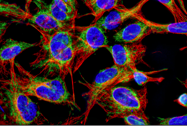
A friend asked when I got a confocal microscope. I will take that as a compliment, I think.
At some point when I have the time, I would like to print some of my diatom photomicrographs and display them in my room. Perhaps I can make them into a calendar or short art book.
Enter to Win IOTY 2022
Whether microphotography is your passion, like Daniel, or your profession, consider submitting to the Evident Image of the Year Award 2022 and sharing your artistic side.
Related Content
Celebrating the Global Image of the Year EMEA Winner’s Festive Fungi
A Growing Art—Meet the IOTY 2021 Americas Regional Winner
All the Fall Things—Our Most Popular Microscope Images for September 2022
