Not Available in Your Country
Sorry, this page is not
available in your country.
Overview
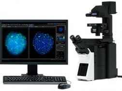
 | The Olympus IX73 inverted microscope has been integrated into our IXplore systems. IXplore Systems are designed to provide solutions-based packages that suit your research application needs. |
|---|
Expandable to Meet Growing Research NeedsThe semi-motorized IX73 is designed to satisfy a variety of research needs. With two deck options and additional modules for expanded functionality, the IX73 is perfectly suited for a changing research environment. |
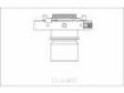
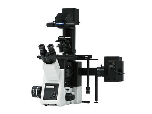
IX73: Two-deck System
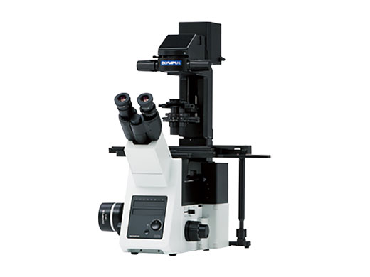
The modular IX73 two-deck system can be combined with coded or motorized units for maximum expandability. | IX73: One-deck System
A microscope designed with an emphasis on efficiency. Ideal for documentation, routine testing and other tasks. |
Need assistance? |
Specifications
| Observation Method > Fluorescence (Blue/Green Excitation) | ✓ | |
|---|---|---|
| Observation Method > Fluorescence (Ultraviolet Excitation) | ✓ | |
| Observation Method > Differential Interference Contrast (DIC) | ✓ | |
| Observation Method > Phase Contrast | ✓ | |
| Observation Method > Brightfield | ✓ | |
| Observation Tubes > Widefield (FN 22) > Tilting Binocular | ✓ | |
| Observation Tubes > Widefield (FN 22) > Trinocular | ✓ | |
| Stage > Manual > Plain Stage | ✓ | |
| Stage > Mechanical > IX3-SVR Mechanical Stage with Right Handle |
| |
| Condenser > Motorized > Universal Condenser | NA 0.55/ W.D. 26.2mm | |
| Condenser > Manual > Universal Condenser | Dry: NA 0.9/ W.D. 1.5 mm, Oil: NA 1.4/ W.D. 0.63 mm (1.25 X - 100 X) | |
| Condenser > Manual > Ultra-Long Working Distance Condenser | NA 0.3/ W.D. 73.3 mm | |
| Dimensions (W × D × H) | 323 (W) x 475 (D) x 656 (H) mm (1 Deck Standard Configuration) | |
| Weight | 35 kg (1 Deck Standard Configuration) |
Related Components
Components
- Spatial and Temporal Precision
- Perfect Reproducibility
- Flexibility
- Cell surface imaging with high Z-resolution
- Minimal background noise
- Wide choice of excitation wavelength
- True multi-color TIRFM
- Software adjustment of penetration depths
- Ease of use
- Perfect control of all environmental conditions
- First complete thought-out workflow-oriented incubation solution
- Open access
Color Cameras
Monochrome Cameras
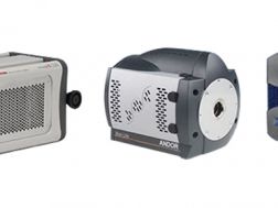
Electron multiplying CCD cameras amplify optical signals without increasing noise. They can capture high-speed images of dim specimens and perform well for fluorescence live-cell imaging with reduced intensity excitation light. They are used to observe protein interaction within a cell and for real-time imaging.
- CCD technology for detecting ultra-low light
- Dedicated, ultrasensitive scientific camera platform
- Quantifiable and reproducible imaging
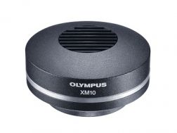
- 1.4-megapixel monochrome cooled CCD
- Low noise and high sensitivity
- Fast Live image speed
Software
Providing intuitive operations and a seamless workflow, cellSens software’s user interface is customizable so you control the layout. Offered in a range of packages, cellSens software provides a variety of features optimized for your specific imaging needs. Its Graphic Experiment Manager and Well Navigator features facilitate 5D image acquisition. Achieve improved resolution through TruSight™ deconvolution and share your images using Conference Mode.
- Improve experiment efficiency with TruAI™ deep-learning segmentation analysis, providing label-free nuclei detection and cell counting
- Modular imaging software platform
- Intuitive application-driven user interface
- Broad feature set, ranging from simple snapshot to advanced multidimensional real-time experiments
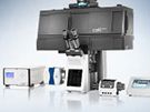
Advanced Imaging Solutions (systems)
Light Sources

Facilitating fluorescence applications, this light-guide-coupled illumination system reduces thermal effects and vibration. It is easy to install and delivers excellent long- and short-term stability. A six-step iris effectively controls intensity and enables simple, staged intensity adjustment, and its pre-centered burner reduces running costs.
- Powerful 130 W burner with long lifetime
- Stable and uniform light output
- Even alignment-free illumination
- Incorporates up to four high power solid state LEDs
- 365, 385, 405, 447, 460, 525, 635, 660 and 735 nm available
- Ultra-fast switching times in microsecond range