Speaking with Dr. Peter Ertl, professor and head of the cell-chip research team at the Technical University of Vienna, (TU Wien) Austria, I learned some fascinating facts about his work with lab-on-a-chip technologies. These tiny self-contained devices can be used to study everything from anticancer drug toxicity to the permeability of the blood-brain-barrier. Now, this technology is being used to develop a COVID-19 diagnostic test that has the potential to rapidly deliver accurate results in just a few minutes.
For Dr. Ertl, the current COVID-19 diagnostic methods lack the combination of speed, sensitivity, and reliability that is needed to properly address the current pandemic. “On one hand, PCR can detect everything, but it’s not scalable and typically takes up to 24 hours to get a result. The result is only really valid for a few days—meaning you can’t go to long-term events, such as conferences. Current alternative methods, such as lateral flow and the antigen testing, are simply not sensitive enough. While they may give good results for symptomatic people with high viral loads, it is equally important to detect asymptomatic people who could be spreading the disease without knowing.”
Developing an Effective, Efficient COVID-19 Diagnostic Tool Using Data-Streaming-Capable Biochips
Tackling these issues head on, Dr. Ertl and his team have developed a biochip technology that they say can reliably detect as few as 3–5 viral particles in just a few minutes while eliminating false negatives. Consequently, asymptomatic people can be definitively diagnosed, enabling them to self-isolate to curb the spread of COVID-19.
The biochip has been designed in such a way that it has its own power supply, display unit, and ability to transfer data wirelessly (Figure 1). Results can be directly and immediately transferred to the necessary epidemiological center. This streamlining of data collection could significantly help in reporting accurate case numbers and outbreaks.
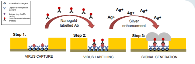
Figure 1. Illustration of how the COVID-19 biochip works. The biochip technology uses an antibody lawn that is immobilized on the microfluidic chamber. These bind any target virus that is present in the sample, in this case the SARS-CoV 2 virus, which causes COVID-19. Subsequently, a secondary nanogold-labeled antibody binds to the bound viral particles. By adding a silver solution at this stage, the silver reacts with the gold to produce conductive nanobridges. In turn, an electrical current can flow through and activate LEDs or even link to an in-built communication device to transfer the data wirelessly.
Role of Olympus’ IXplore™ Live Cell Imaging Platform in Validating the COVID-19 Biochip Test
The proper functioning of the technology is contingent on the quality of the antibodies and how well they are immobilized onto the microfluidic chamber surface and spacing between the interrupted electrical leads. To optimize this, Dr. Ertl and the cell-chip research team tested different immobilization strategies. Using the Olympus IXplore™ Live microscope system and secondary fluorescently labelled antibodies, they were able to assess key parameters—including antibody density, orientation, and alignment—that were critical to the success of the biochip (see Figure 2).
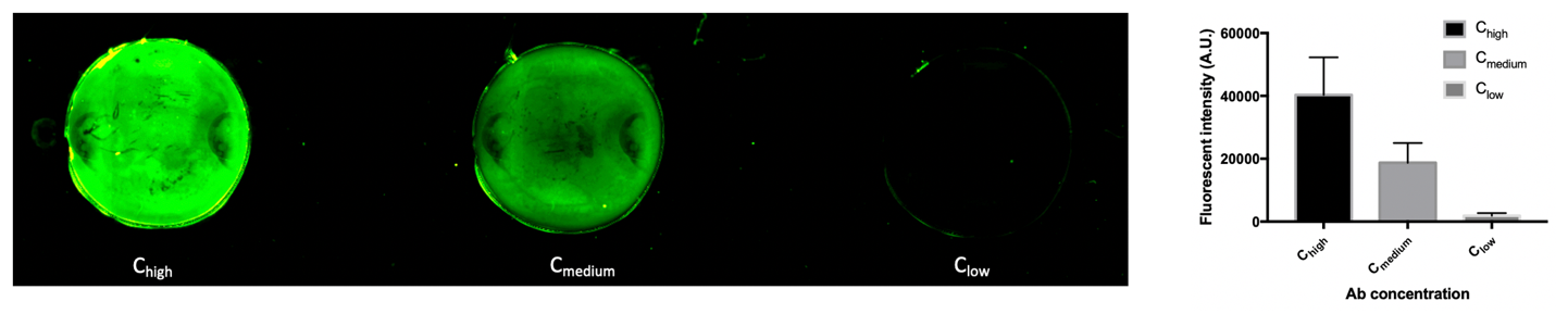
Figure 2. Using the Olympus IXplore Live microscope and secondary fluorescently labeled antibodies to determine the immobilization of a goat-rat IgG antibody density on glass substrate at 4x magnification. The image shows decreasing levels of antibody concentration (from left to right) in line with decreasing levels of fluorescence intensity.
Dr. Ertl elaborated, “The IXplore Live system has been really useful to augment chip development—it offers our lab exactly what we need in terms of image resolution and price. Outside of COVID-19, it’s become the true workhorse of our lab, being used perpetually for our organ-on-a-chip projects. Particularly, the excellent resolution, the hypoxic chamber, and the ability to upgrade the system to confocal imaging has been extremely beneficial to our research.”
Applications for Other Diseases, Including Cancer and Parkinson’s
Aside from the viral diagnostic platform, the biochip technology is being harnessed for a variety of exciting applications. In Dr. Ertl's lab, they are researching advanced microfluidic cell culture systems that can replicate the complex 3D architecture of tissues and organs, known as organ- or tissue-on-a-chip devices. Not only are these biochips being used as biological models to investigate structure and function of tissues, but they are also providing insight into the onset and progression of diseases such as cancer, autoimmune, and neurodegenerative diseases.
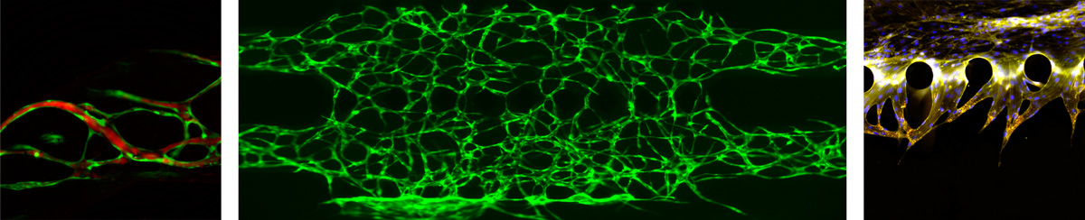
Figure 3. Images taken by the research team at TU Wein using the Olympus IXplore Live microscope. From left to right the images show (1) GFP labelled HUVECs with TRITC-dextran perfusion (20x magnification) (2) GFP labelled HUVECs (4x magnification) (3) DAPI labelled F-actin and VE-Cadherin (20x magnification).
Dr. Ertl explained that the IXplore Live microscope has become an important tool for organ-on-a-chip research. The high resolution is essential to validate 3D cell assembly—making sure that the architecture mimics that of actual human tissue structure. For example, the team at TU Wien have used the IXplore Live microscope to follow the progression of the lymphatic/blood interface as well as the development of a sensor-integrated Parkinson’s disease on a chip platform. In the latter project, the IXplore Live microscope has been central to studying neurite outgrowth and calcium imaging. The research team are hoping this will lead to a personalized human midbrain model that will advance our understanding of the neurodevelopmental aspects of Parkinson’s disease.
Additionally, the group noted that the ability to combine the IXplore Live microscope with a hypoxic chamber was a critical choice for their lab—being involved in the development of nearly all microfluidic devices. Sarah Spitz, a member of Dr. Ertl’s research group, commented “We mainly use the hypoxic chamber to study oxygen permeability of various materials for the fabrication of microfluidic devices. Oxygen permeability is an important parameter as it will affect the availability of oxygen for the cells that are grown. Using this setup and an integrated oxygen sensor within the chip, we can determine these characteristic properties very easily.”
Potential of Biochip Technology Beyond COVID-19
Viral testing on biochips not only represent a potentially rapid, reliable tool to monitor the ongoing pandemic, but investment in the platform could be fruitful for diagnosis of other diseases. Being self-powered and portable, the technology could be harnessed in remote areas or where vaccine storage is an issue, such as for Hepatitis B in Pakistan and Ebola in Africa. Equally, the future of organ-on-a-chip technology is full of potential and promises exciting developments in the treatment of many diseases. As for the COVID-19 test, Professor Ertl says that they hope to develop the first industrial prototype within the next few months.
Related Content
Microscopy Meets Microfluidics—Creating an Adaptable Imaging System for Modern Research
Remote Microscopy Guide: 6 Tips to Set Up Your Lab for Success