As a leading microscope manufacturer, Olympus provides various optical components designed to integrate into microscope-based imaging systems used in a wide range of fields, including life science research, medicine, and industries such as manufacturing.
To support this effort, we have been researching and developing our own illumination optical systems. The result is the integration of Olympus’ innovative True Color LED into our U LHLEDC and U LHLEDC100 lamps. The True Color LED is a bright light source that has a high intensity and long life. Most importantly, it features spectral properties that closely match those of conventional halogen lamps. These powerful and efficient illumination systems are offered as part of our OEM optical components.
Halogen Lamps versus LEDs for Microscopy
Conventional halogen lamps have been used for many years as the gold standard of microscope light sources because of their excellent color-rendering abilities (both through the eyepiece and on computer monitors). Thanks to advances in LED technology, bright and efficient LED lighting has become an increasingly popular option at both home and work. However, in the microscopy field where the light source’s ability to accurately render color can significantly affect the results of an evaluation, LED technology historically had a disadvantage. The spectral uniformity of LEDs could not match the performance of conventional halogen lamps, meaning the color reproduction was unreliable. This shortcoming made it difficult to integrate generic white LEDs into microscopes.
Introducing the True Color LED into Our OEM Optical Components
Our engineering team tackled this problem by developing the True Color LED and integrating it into our illumination systems (U-LHLEDC and U-LHLEDC100). This light source provides high color rendering without compromising the advantages of LEDs:
- Low energy consumption
- Long lifespan
- Bright, uniform illumination
Our True Color LED has overcome the drawbacks of generic white LED light sources with its ability to accurately render color. Substantial improvement in spectral properties has resulted in color reproduction that closely matches that of halogen lamp illumination. See how close in the graph below.
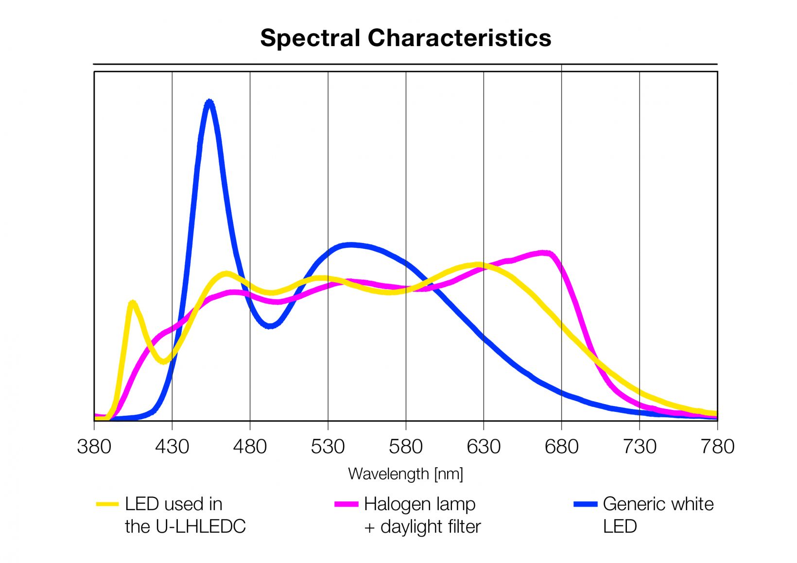
The spectral characteristics of the True Color LED used in the U-LHLEDC illumination system (yellow), a halogen lamp with a daylight filter (pink), and a generic white LED (blue). The True Color LED shows similar performance to halogen.
As a result, our U-LHLEDC and U-LHLEDC100 illumination systems benefit from the high color rendering capabilities of the True Color LED. These systems have already been adopted as optical components in many devices for pathological and cytological use in the life science field and in Raman spectrometers in the industrial sector.
In pathological examinations, our LED technology enabled pathologists to compare samples against large amounts of data on cancer cells acquired through observation with halogen lamps. The True Color LED’s proven performance in these experiments paved the way to its use in clinical studies performed on cancer patients.
Check out our white paper on the True Color LED to read more about this innovation.
To learn more about our high-quality optical components and parts that integrate into microscope designs, visit our online resource center for OEM microscope components.
Related Content
White Paper: Trust the Colors with Olympus True Color LED
OEM Microscope Components for Integration
A Faster Way to Verify RoHS Compliance of Microscope Components and Optics