
In July, our most popular image was, quite literally, the star of the show. We’re thrilled each year when it’s time to announce the winners of our global scientific imaging competition, and seeing the winning images in our top image roundups always makes us happy. This month also boasts a beautiful selection of plants and animals, from spinach to cats and dogs.
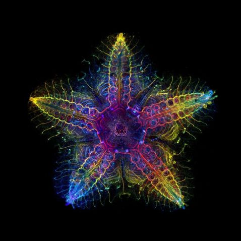
It’s no surprise that our most popular image this month is this stunning image of the nervous system of a juvenile sea star (Patiria miniata). Captured by Laurent Formery from the United States, this image was selected as the global winner of our 2022 Image of the Year (IOTY) competition. The small sample, about 1 cm wide, was labeled with an antibody against acetylated tubulin after optical clearing and
captured using a color-coded Z-projection. Laurent chose to image this sea star to showcase the beauty of science in the oceans.
“I work with marine invertebrates, in particular echinoderms (sea stars, sea urchins and their kind),” commented Laurent. “They are beautiful animals, with a fascinating and aesthetically pleasing fivefold symmetry that is unlike anything else in the animal kingdom. We know little about how these animals shape their fivefold body, which is the topic of my research. Echinoderms, and marine invertebrates in general, are often not well-known animals. I’m happy that taking
images of them helps communicate how much beauty we have in our oceans, and why it is important to know more about them and protect them.”
Image courtesy of Laurent Formery, global winner of the 2022 IOTY competition. To see all the winning images, go to olympus-lifescience.com/ioty.
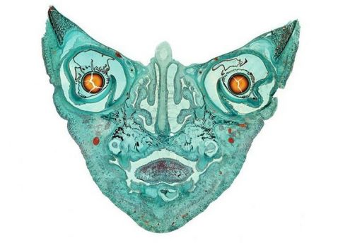
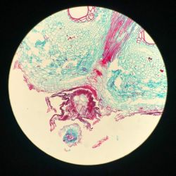
This image is an old favorite and, apparently, we aren’t the only ones who love when it reappears! Here we're looking at an amazing cross-section of a fetal cat. Or is it looking at us? The microscope image shows maturing ears, eyes, tongue, skull bone, sinus cavities, and other skull features. The sample was 12 mm in diameter.
This stunning image was captured after the fetal cat skull was prepared for examination and cut in a longitudinal view. Four individual images were combined into one final file called a computational photograph, which offers enhanced resolution and image enlargement.
Image courtesy of Mike Peres. Captured using brightfield microscopy at 2X magnification.
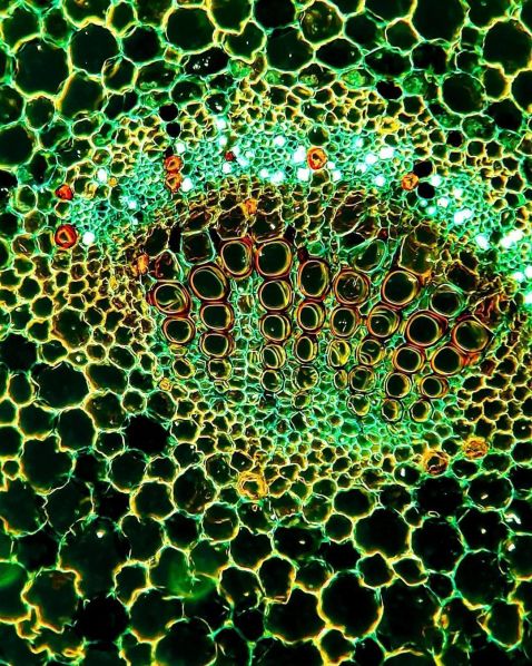
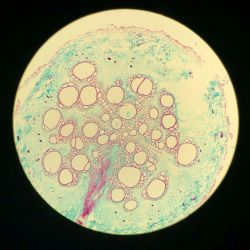
Plants under the microscope never fail to amaze us!
"Dicotyledon plants have distinguishing nodes and internodes, giving rise to leaves, branches, and flowers, contrary to monocotyledon plants. The inner structure of vascular bundles consists of xylem (marked as orange and yellow), phloem (marked as fluorescent green), and multiple fibers."
Image and caption courtesy of Minjun Shin. Captured using an Olympus CX21 microscope.
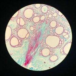
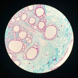
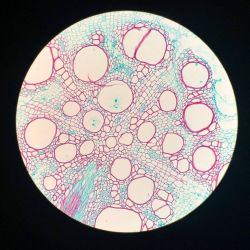
Maybe Popeye was on to something, because just looking at these images of spinach gives us a boost of energy. We’ll take a whole bowlful of these beautiful images, please! This stunning series of firework-like images captures a spinach root cross-section.
Image courtesy of UiM. Captured using an Olympus BH2 microscope.
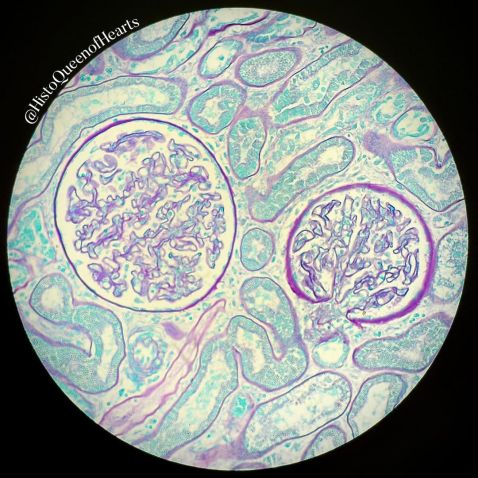
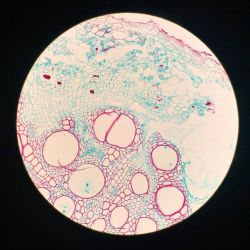
We must have antioxidants on our minds, because this pretty image reminds us of the inside of a pomegranate. Don’t be fooled though: it’s actually a dog’s kidney!
"Made with Periodic acid–Schiff (PAS) with a light green counterstain, this image shows two beautiful glomeruli. The glomeruli in kidneys are made up of many little capillary loops and bundles. This is where the filtration process of the blood stream begins. These capillary bundles are lined by something called a basement membrane. This membrane has an affinity for the Schiff reagent, so it stains bright pink."
Image and caption courtesy of Kate Murphy. Captured using an Olympus BX40 microscope.
Bonus video! Chloé Savard is back with our favorite specimen—tardigrades!
"These cuties are among my favorite tardigrades: Hypsibius sp. I keep a culture at home, and they’re starting to make babies. You can see a couple in this video!
The word tardigrade was chosen by an Italian biologist. It literally means ‘slow steppers’ in Latin, which is pretty funny because some of them are pretty fast on their eight feet, in my opinion!
Tardigrades, also called water bears or moss piglets, are without a doubt the international super stars of the microworld. They’re famous because they evolved and adapted to survive extreme environmental conditions. Among these adaptations are resisting dehydration by forming a quiescent tun and resisting sub-zero temperatures down to -196 °C (-320.8 °F) in their hydrated form and temperatures from -273 °C to 100 °C (-459.4 °F to 212 °F) in their
dehydrated form. They can also survive an enormous amount of radiation, high pressure, low oxygen concentration, and even exposure to the vacuum of space. It has been shown that water bears are among the most radiation-tolerant animals on this planet!
All of these adaptations allowed tardigrades to inhabit every microenvironment on Earth; from Arctic to Antarctic, deserts to tundra, mountains and forests, grasslands and valleys, ponds and lakes. They can even be found in the deep sea and most likely in your own garden! Since they usually measure between 50 microns to 1.2 millimeters, using a microscope to observe those from your garden will be necessary.
Can you spot the bubble-like structures moving around the water bear’s body?"
Video and caption courtesy of Chloé Savard. Captured using an Olympus BX53 microscope. Check out all of Chloé’s takeover posts on the Evident Life Science Instagram!
To see more images like these, be sure to follow us on Instagram at @evidentlifescience!
Want to share your own images? Visit our image submission site.
Related Content
Easy Being Green—Our Most Popular Microscope Images for June 2023
Salt to Stinging Nettles—Our Most Popular Microscope Images for May 2023
Hibiscus to Vanilla—Our Most Popular Microscope Images for April 2023
