
When Kermit the Frog lamented that “it’s not easy being green,” he must not have seen our latest Instagram content! Shades of bold greens are seen throughout our most popular microscope images for June, from brine shrimp and moss to summertime beverages.
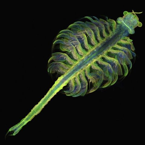
We love when old favorites continue to impress! Commonly known as brine shrimp, this is a stunning image of Artemia, a genus of aquatic crustaceans. Able to live in bodies of water with up to 25% salinity, these microorganisms are commonly found in inland saltwater lakes.
Image courtesy of Gopi Shah, an honorable mention in our 2018 Image of the Year (IOTY) competition.
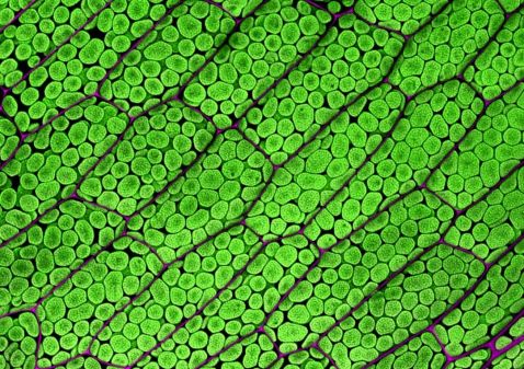
“Here is a cool fluorescent image of moss gametophore ‘leaf’ cells. In the image, we can see chloroplasts (in green) and walls between neighboring cells (in magenta). Both are visible due to their natural autofluorescence. This is a deconvolved maximum Z-projection image. While the image shows the maximum projection, a video [shown on Ivan’s Instagram] portrays the 3D reconstruction of the Z-stack, which shows that only a small section of the cells was captured in the Z-stack.”
Image and caption courtesy of Ivan Radin, the Americas regional winner of our 2021 IOTY competition. Captured using an Olympus FLUOVIEW™ FV3000 confocal microscope. To learn more about Ivan, check out our interview about his winning IOTY image.
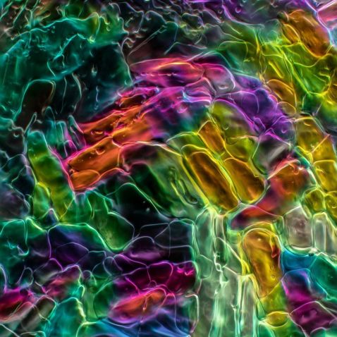
The hot summer sun means it’s time to break out the spritzes and lemonades by the pool. We know that gin and lemonade are a refreshing combination, but we had no idea how much beauty lay beneath the surface!
Image courtesy of Karl Gaff. Captured with an Olympus X Line™ objective.
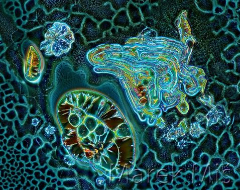
The pretty blue and green hues in this image make us think of swimming in the ocean. While this image may look like an abstract painting of a coral reef, it’s actually a micrograph of a recrystallized mixture of hydroquinone (a pigment lightener) and erythritol (a sweetening agent) captured under polarized light.
Image courtesy of Marek Miś. Captured using an Olympus BH2 microscope.
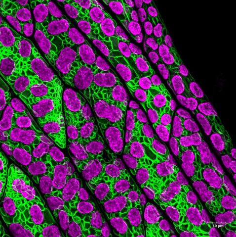
“A few cells from a ‘leaf’ or phyllid from moss Physcomitrium patens gametophore. Cells are expressing a green fluorescence marker that is labeling the vacuolar membrane. Chloroplasts are also visible (in magenta) due to the autofluorescence of chlorophyll. Fun fact: Moss phyllids are only one cell layer thick in some regions.”
Image and caption part of an Instagram takeover courtesy of Ivan Radin. Captured using an Olympus FV3000 confocal microscope with a 60X water immersion objective (1.2 NA).
Our bonus video shows snack time for this little rotifer!
"This weird-looking creature is giving some extraterrestrial vibes, but it’s actually among the smallest animals on Earth! It’s called a rotifer, and this particular species was named Stephanoceros fimbriatus. Could’ve been a dinosaur name, in my opinion. In Greek, 'Stephano' means crown and 'ceros' means horn, which makes total sense when looking at this microscopic monster.
Rotifers are also referred to as ‘wheel animals’ because of the rotating cilia, called corona, on their heads! They usually use these cilia to create a water current that attracts food particles like bacteria, algae, ciliates, and other microorganisms to the mouth. Although this species is a bit different from the other rotifers.
Instead of having some rotating wheels on their heads, they have five long tentacles with moving cilia that redirect prey toward the mouth! These rotifers are basically ambush predators waiting for prey to pass by. The prey then get trapped into small compartments before getting pumped by sphincter muscles and ultimately being swallowed."
Video and caption courtesy of Chloé Savard. Captured using an Olympus BX53 microscope.
To see more images like these, be sure to follow us on Instagram at @olympuslifescience!
Want to share your own images? Visit our image submission site.
Related Content
Salt to Stinging Nettles—Our Most Popular Microscope Images for May 2023
Hibiscus to Vanilla—Our Most Popular Microscope Images for April 2023
Colors of Spring—Our Most Popular Microscope Images for March 2023
