
From eukaryotic organisms and leaves to kidneys, our most popular images for August 2021 showcase a rainbow of colors. View the colorful captures below!
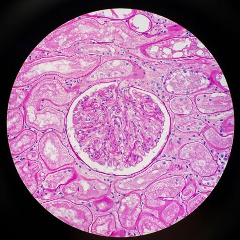
Seen here is a section of the kidney from a tiger stained with Periodic acid–Schiff (PAS).
“The purpose of kidneys is to filter the blood. This is where waste is removed and collected to excrete in urine. The kidneys are also responsible for regulating the amount of salt, water, and minerals in our bloodstream.
The glomerulus, in my opinion, is the star of the show. It’s a loop of capillaries within the kidney, and it’s where the filtration process begins. The small molecules like water and wastes will filter out and travel down to the tubules. As it travels down the tubule, important substances are then returned back into the bloodstream, leaving the wastes to travel into a collecting duct and eventually be excreted in the urine.”
Image and description courtesy of Kate Murphy. Captured on an Olympus BX40 microscope.
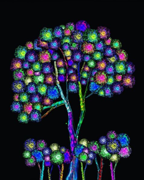
Appropriately titled "Tree of Nerve," this microscope image is not a plant or flower but actually a collection of nerves. The flowers are made from a dorsal root ganglion cluster and Schwann cells explant cultures, while the branches and trunks are made from sciatic nerves stained for Schwann cells and neurofilament.
Image courtesy of Diara Santiago Gonzalez. Captured on an Olympus disk scanning unit (DSU).
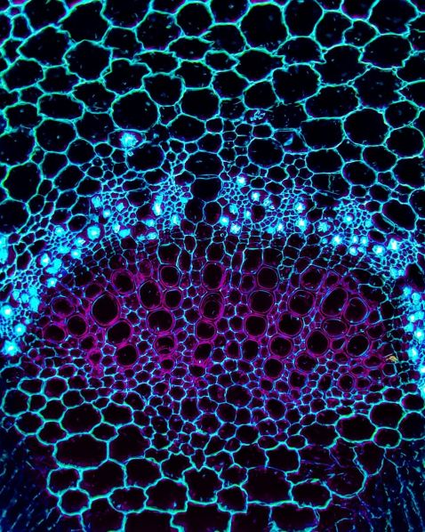
Our next image is a magnified look at a cotton leaf, captured using a custom blue Rheinberg filter. Rheinberg illumination, a form of optical staining, is a striking variation of low to medium power darkfield illumination using colored gelatin or glass filters to provide rich color to both the specimen and background.
The cross section of the cotton leaf reveals a complex meshwork-like structure that consists of xylem (purple), phloem (light blue), and multiple fibers.
Even more incredible than the image itself is that it was captured by a high school biologist and photographer from South Korea. We love to see young scientists at work!
Image courtesy of @minjunsmicroworld. Captured on an Olympus CX21 microscope.
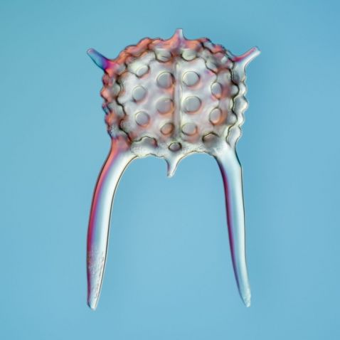
As part of our "Exploring the Microscopic World with X Line™ Objectives" series with Håkan Kvarnström, each month we share a new image showing the microscopic life of the ponds in and around the Stockholm Royal Gardens. This month's featured image shows the intriguing shape of the skeleton of a Radiolarian, captured using DIC observation and focus stacking (20X). These skeletal remains produce siliceous ooze, a type of sediment that makes up 15% of the ocean floor!
Image courtesy of Håkan Kvarnström. Captured on an Olympus BX51 microscope with X Line objectives.
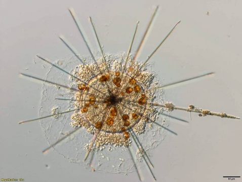
Acantharia are a protist (an organism whose cells contain a cell nucleus that is not an animal, plant, or fungus) that can frequently contain symbiotic algae. In this close relationship between algae and protist, specialized nutrient cycling can develop. The shape and size make Acantharia easy to identify within plankton tows. Their skeletons are made of strontium sulfate (SrSO4). They are one of the only organisms that are primarily made of this sulfate salt!
Image courtesy of Gabrielle Corradino. To learn more about her work, check out our Discovery Blog interview.
Bonus video!
What you're watching is a leaf during photosynthesis at full throttle. Look closely to see the disk-shaped chloroplasts traveling around the cells in a regular circulating pattern, almost taking the same path at each turn. This is made possible by protein machines that work like conveyor belts to pull the green organelles around and expose them evenly to light.
Video courtesy of Benedikt Pleyer.
To see more images like these, be sure to follow us on Instagram at @olympuslifescience!
Interested in sharing your own images? Visit our image submission site.
Related Content
Embracing Social Media and Its Important Role in Science Outreach
Avalon Finds Her Dream Microscope to Study Rotifers
Body Part Art: Our Most Popular Microscope Images for July 2021
