
July’s most popular images were all about body parts. From the brain to the spleen to the eyeball, we’ve had some close looks at what makes up a body.
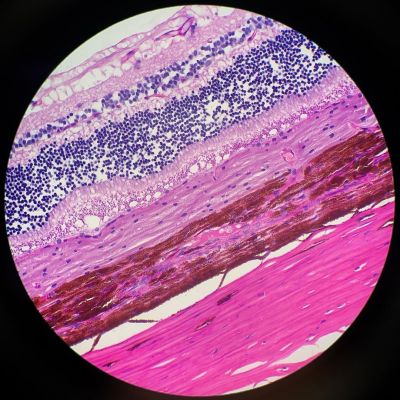
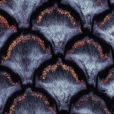
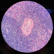
Kate Murphy’s stunning pink stripes aren’t simply a pretty pattern, but an image of a section of tissue from a dog’s retina.
Kate’s enthusiasm for her subject is clear:
“The retina is a very thin layer of specialized cells at the back of the eyeball, where the optic nerve is. The retina receives light and converts it into neural signals. The signals are then sent to the brain for visual recognition.
The retina is comprised of ten layers:
- Retinal pigment epithelium
- Photoreceptors
- External limiting membrane
- Outer nuclear layer
- Outer plexiform layer
- Inner nuclear layer
- Inner plexiform layer
- Ganglion cell layer
- Nerve fiber layer
- Inner limiting membrane
I love eyeballs! They are truly such a fascinating organ. It’s crazy to think that without that little layer of cells, we wouldn’t be able to see!”
Image and description courtesy of Kate Murphy. Captured on an Olympus BX40 microscope.
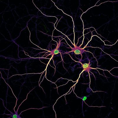
Cultured neurons are a good model to test enzyme replacement therapy (ERT) for many neurological disorders. This striking image was collected from an experiment conducted by researcher Nadia Efimova, where she studied the uptake of engineered enzymes in rat cortical neurons (staining of the enzyme is not shown). The image shows primary rat cortical neurons at 14 days in vitro with nuclei (green) and developing dendrites (mpl-inferno LUT). MAP2 staining was added to visualize outlines of neurons.
Image courtesy of Nadia Efimova, Olympus 2020 Image of the Year Award Honorable Mention.
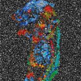
To celebrate World Emoji Day on July 17, we transformed a few of our winning 2020 Global Image of the Year images into emojified works of art! Starting at the top left:
Anther of Arabidopsis arenosa.
Image courtesy of Jan Martinek, 2020 Image of the Year Award Honorable Mention.
Collagen fibers and dermal pigment cells in African house snake embryonic skin.
Image courtesy of Grigorii Timin, 2020 Image of the Year Award Europe, the Middle East, and Africa Regional Winner.
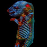
Whole rat embryo.
Image courtesy of Werner Zuschratter, 2020 Image of the Year Award Global Winner.
Scales collected from the wings of over 40 species of butterflies.
Image courtesy of XinPei Zhang, 2020 Image of the Year Award Asia-Pacific Regional Winner.
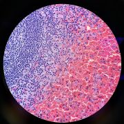
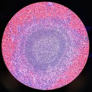
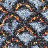
Kate Murphy’s beautiful histo stains made the top five posts again with these stunning spleen images.
“The spleen is the largest organ in our lymphatic system. It stores white blood cells and recycles old red blood cells. It’s basically the blood’s quality control center.
Microscopically, we can see the two different “pulps.” White pulp is the purple, circular clusters of cells. This is the lymphoid tissue. Red pulp (filled with red blood cells) is where blood filtration occurs.
This spleen is from a red-necked wallaby.”
Images and description courtesy of Kate Murphy. Captured on an Olympus BX40 microscope.
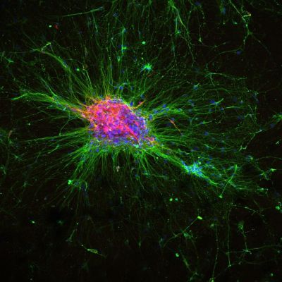
This image shows an iPSC-derived “organoid” differentiated to produce dopaminergic neurons. This experiment aims to model rare neurodevelopmental diseases using these cells and CRISPR-Cas9 gene editing techniques.
Image courtesy of Scott Bell from the Olympus Discovery Center at the Douglas Institute at McGill University, part of a program designed to help advance bioscience by making advanced microscopy equipment available to researchers.
To see more images like these, be sure to follow us on Instagram at @olympuslifescience!
Interested in sharing your own images? Visit our image submission site.
Related Content
Bugs and Brains: Our Most Popular Microscope Images for May 2021
Avalon Finds Her Dream Microscope to Study Rotifers
Oceans, Organs, and the Outdoors: Our Most Popular Microscope Images for June 2021
