
Another month of incredible microscope images, and another series of alliterative samples. This month, the most popular images include brains, bugs, and bacteria!
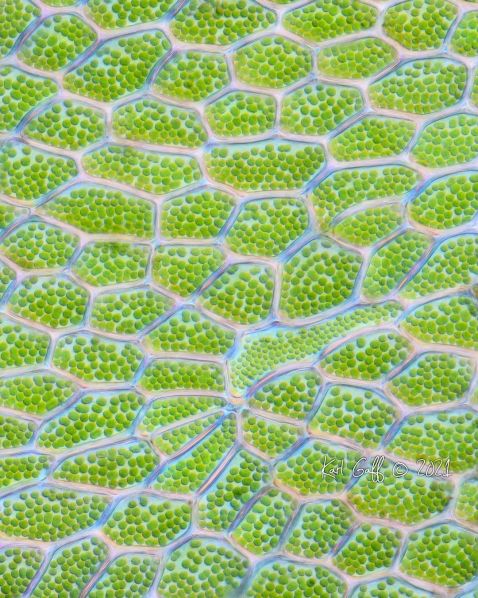
The most popular image of the month is simple but stunning. This close-up shows the fine details of chloroplast-filled polygonal cells from moss collected by microscopist Karl Gaff in the Wicklow Mountains, located just south of Dublin, Ireland.
Image courtesy of Karl Gaff. Captured using DIC focus stacking with an Olympus 2X / 0.80 X Line objective.
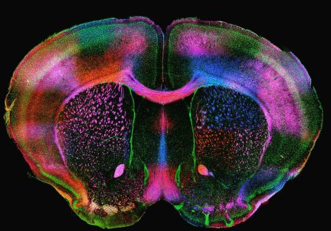
All brains are beautiful, but this "rainbow brain" is truly one-of-a-kind! This beautiful image of a mouse brain is expressing myelin proteins PLP (proteolipid protein) and MBP (myelin basic protein) in multiple colors. Myelin is a fatty substance that surrounds nerve cell axons and acts as insulation. The proteins not only look beautiful, but also work to maintain the structure of the myelin.
Image courtesy of Diara Santiago González. Captured on an Olympus disk scanning unit (DSU).
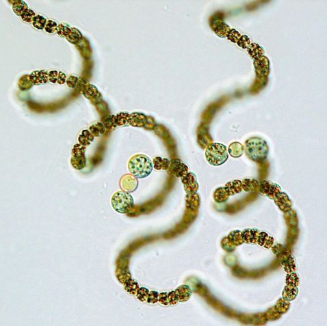
Planktonic cyanobacteria are a fundamental component of marine food webs and are major contributors to global carbon and nitrogen fluxes. This common type, Sphaerospermopsis reniformis, may be difficult to try to pronounce, but is easy to identify under the microscope. It’s characterized by spherical, thick-walled cells known as akinetes that are always adjacent to the heterocytes, which are nitrogen-fixing cells.
Image courtesy of Glenn McGregor.
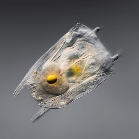
Rotifers are truly the definition of small but mighty.
“This is a rotifer Polyarthra. Rotifers are the smallest animals on earth. This one is only 150 micrometers long (0.15 mm). This is a female. We can clearly see 5 nuclei in the vitellarium (a modified part of the ovary) and a yellow yolk. Rotifers have existed for at least 35 million years as they have been identified in fossilized amber. They only live for about 1–2 weeks. Under tough conditions (e.g., drought), rotifers can go into a desiccated state as a cyst or egg that can survive for decades without any signs of aging. The cysts can be carried by the wind and allow the rotifer to spread and escape harmful conditions. Once it finds water, the rotifers come back to life!”
Image and description courtesy of Håkan Kvarnström. Captured on an Olympus BX51 microscope with X Line objectives.
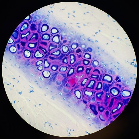
Kate Murphy, the “histology queen of hearts” has been sharing images each week for her “Special Stain Saturday.” Follow her to learn staining tips and techniques and see more striking images like this one.
“Ever wondered how a certain tissue or structure would react to a stain? I always do!! I love experimenting with specials and seeing how the different tissues react. And I’ve encountered some pretty amazing results. Today’s stain is AFB (acid fast bacteria). Typically, it’s used to identify acid-fast bacteria, but recently I discovered that it has the most beautiful effect on cartilage! AFB is starting to become one of my new favorite stains to test out on tissues.”
Image and description courtesy of Kate Murphy. Section of elastic ear cartilage captured on an Olympus BX40 microscope.
Back by popular demand, Benedikt Pleyer took over our channel for another exclusive Instagram Takeover.
“It is fascinating to see how a mosquito gets its blood meal. The proboscis consists of a labia part wrapped around the needle. When the mosquito stings, the labia hold the needle in place like an oil rig holds the drill during a fracking operation. And this is what the mosquito does quite literally. It injects saliva while drilling into blood-rich capillaries.”
Video and description courtesy of Benedikt Pleyer. Captured with an Olympus MPlan objective at 2.5x magnification with darkfield.
To see more images like these, be sure to follow us on Instagram at @olympuslifescience!
Interested in sharing your own images? Visit our image submission site.
Related Content
Snakeskin on a Microscopic Scale: Presenting the IOTY 2020 EMEA Regional Winner
Protists, Plants, and Tigers: Our Most Popular Microscope Images for August 2021
Embracing Social Media and Its Important Role in Science Outreach
