
Advance Your Research with 4 Innovative Technologies for FLUOVIEW
- TruSpectral: High light efficiency for bright, precise multicolor images.
- TruSight: Seamless deconvolution for clearer, sharper images.
- TruFocus: Stable time-lapse images with low phototoxicity.
- TruResolution: Automated correction for brighter and sharper images at depth.
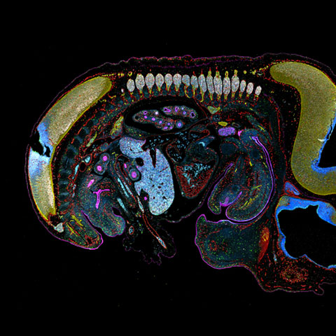
Brainbow AAV transfection of Purkinje cells amplified with antibodies as described in Cai et al 2013. Visible are Purkinje cell somata, dendrites and axons, as well as some aspecific stainings of granule cells. | TruSpectral - High Sensitivity Multi-Color Imaging(Compatible with FV3000)An integrated feature of all FLUOVIEW FV3000 confocal systems, TruSpectral technology enables much higher light throughput compared to conventional spectral detection units. TruSpectral technology employs volume phase holograms that diffract light more efficiently than traditional reflection gratings, delivering up to three-fold higher transmission for red and far-red wavelengths. The result is excellent multicolor fluorescence with low excitation light for live and fixed tissue imaging. |
|---|
TruSight - GPU Based Deconvolution
(Compatible with FV3000, FVMPE-RS)
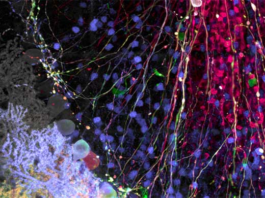
Remove blur and obtain clearer, sharper images with TruSight deconvolution. Specialized cellSens algorithms for the FLUOVIEW FV3000 confocal microscope and FVMPE-RS multiphoton microscope enable a seamless workflow from acquisition to publication with the click of a button.
 HeLa cells Blue: nuclei (DAPI), Green: microtubules (Alexa Fluor 488), Red: mitochondria (MitoTracker Red) | Nucleopore Nup153 (Alexa Fluor 488), Nup62 (Alexa Fluor 555) Image data courtesy of: Dr. Hidetaka Kosako, Fujii Memorial Institute of Medical Sciences, Tokushima University. |
Related Videos3D imaging NK cells attacking spheroid HT-29 tumor cells for 48 hours. | TruFocus - Stable Time-Lapse Imaging(Compatible with FV3000, FVMPE-RS)The TruFocus system makes multiposition experiments and long-term, time-lapse imaging more robust and reliable. The system uses a minimally phototoxic beam to accurately maintain the focus position, so you don’t have to worry about thermal drift caused by a change in room temperature or drug delivery during experiments. |
|---|
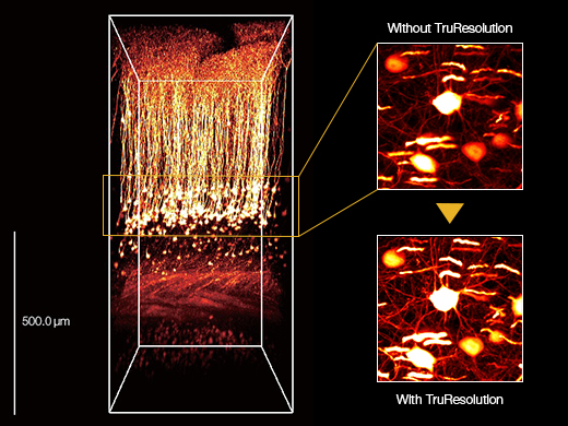
TruResolution - Maximize Resolution in Deep Imaging(Compatible with FVMPE-RS)Maximize the deep imaging performance of your multiphoton microscope with TruResolution objectives. An automated correction collar compensates for spherical aberration at every depth, delivering bright, high-resolution images from the top to bottom of your image stack. |
Maximum projection of mouse in vivo brain images acquired at around 600 um depth (Thy1-YFP-H mouse, sensory cortex). |
|---|
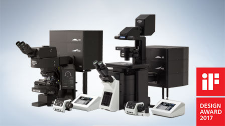
Related Products
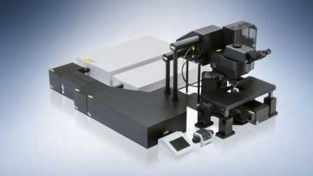
FV3000 Confocal Laser Scanning Microscope
|
|
FVMPE-RS Multiphoton Laser Scanning Microscope
|
|
Sorry, this page is not
available in your country.
Sorry, this page is not
available in your country.