There are two major differences between the mitochondrion and most other organelles. First, it has its own circular DNA (similar in structure to the DNA of prokaryotes). Secondly, it reproduces independently of the cell in which it is found; an apparent case of endosymbiosis. Scientists hypothesize the origins of this circumstance go back millions of years to a time when small, free-living prokaryotes were engulfed, but not consumed, by larger prokaryotes, perhaps because they were able to resist the digestive enzymes of the host organism. The two organisms developed a symbiotic relationship over time, the larger organism providing the smaller with ample nutrients and the smaller organism providing ATP molecules to the larger one. Eventually, according to this view, the larger organism developed into the eukaryotic cell and the smaller organism into the mitochondrion. In the digital video presented above, normal Gray fox lung fibroblast cells (FoLu line) are expressing a fusion of Kindling Red fluorescent protein to a mitochondrial targeting sequence.
Video 1 - Run Time: 06 Seconds
Video 2 - Run Time: 10 Seconds
Video 3 - Run Time: 07 Seconds
Video 4 - Run Time: 07 Seconds
Video 5 - Run Time: 13 Seconds
Video 6 - Run Time: 14 Seconds
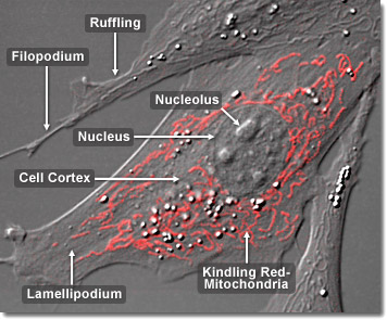
Enzymes of the mitochondrial respiratory chain (also referred to as the electron-transport chain) are embedded within the inner, highly convoluted membrane of all mitochondria. Besides acting as highly selective filters allowing only specialized molecules into the inner matrix, these enzymes are also essential in the animal cell to the production of adenosine triphosphate (ATP) from the diphosphate analog, a process known as oxidative phosphorylation. The generation of the proper amount of ATP necessary to sustain life is related to the number of infoldings of the cristae. More folds produce a larger surface area for the inner membrane, which in turn yields greater amounts of ATP. For example, there are three times as many folds on the inner mitochondrial membrane in heart cells than there are in liver cells because of the larger energy needs of the heart muscle. In the digital video presented above, normal Gray fox lung fibroblast cells (FoLu line) are expressing a fusion of Kindling Red fluorescent protein to a mitochondrial targeting sequence.
Mitochondria appear to be primarily inherited through the maternal lineage in most animal species, though some recent evidence suggests that in rare instances mitochondria may also be inherited via a paternal route. In most cases a sperm carries mitochondria in its tail as a source of sustenance for its long journey to the egg. When the sperm attaches to the egg during fertilization, the tail falls off. As a result, the only mitochondria the offspring acquire are from the egg that its mother provided. Therefore, unlike nuclear DNA, mitochondrial DNA doesn’t get rearranged with every succeeding generation, so it is presumed to change at a much slower rate, giving it high value for the study of human evolution. Mitochondrial DNA is also an important tool in forensic science for identifying corpses or body parts, and has been implicated in a number of genetic diseases, such as Alzheimer’s disease and diabetes. In the digital video presented above, normal Gray fox lung fibroblast cells (FoLu line) are expressing a fusion of Kindling Red fluorescent protein to a mitochondrial targeting sequence.