University of Colorado Boulder
|


Facility Staff |
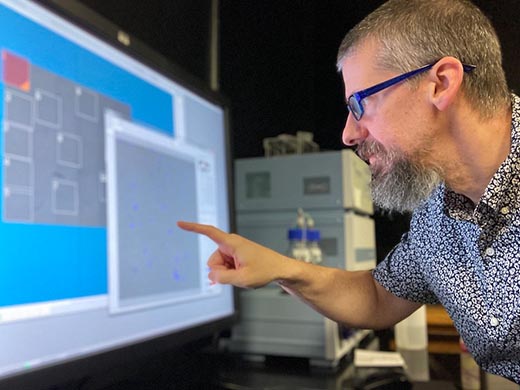
| James D. Orth, PhD, Light Microscopy Core Facility DirectorThroughout his career, Dr. Orth has studied many aspects of cell and molecular biology, including gene regulation, centrosome biology, membrane trafficking, actin and cell motility, and mitosis – using advanced microscopy as a central approach. His own independent research has focused on cancer therapy development. During anticancer drug response, there is often a disconnect in understanding molecular responses in cells and their fates. This is due to profound heterogeneity within the cancer cell population and tremendous variability in drug responses, which is difficult to study directly. Orth developed quantitative, longitudinal microscopy methods to improve our understanding of therapeutic action. The LMCF under Dr. Orth relentlessly helps investigators apply advanced microscopy to help answer many questions in biology, chemistry, physics and engineering that are outlined above. |
 | “Our partnership with Evident will help make advanced microscopy resources available to the entire university. The capabilities afforded through these microscopes will enable important discoveries that will help propel our faculty and students in their careers while also fueling advances for the broader scientific community.”—Joe Dragavon, PhD, Director of Core Facilities and Shared Instrumentation at the University of Colorado Boulder |
In the Spotlight
|

Systems at CU Boulder | |
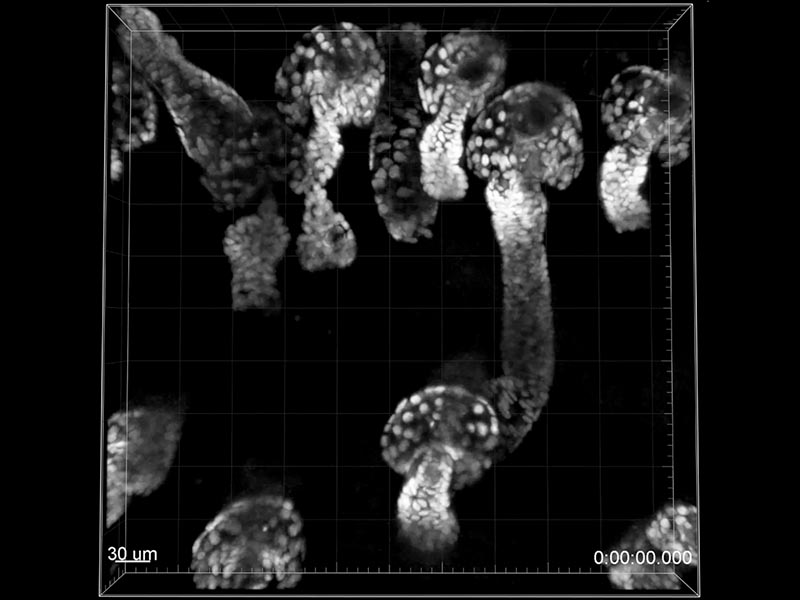
FVMPE-RS multiphoton microscope
Achieving high-sensitivity, high-resolution imaging deep into biological specimens, the FVMPE-RS multiphoton microscope reveals how cells function and interact within living tissue. | IX83 IXplore™ live cell system
Offering enhanced cell viability for physiological experiments, the IXplore live cell system with its integrated IX83 microscope reduces photobleaching and helps researchers maintain their samples under strict physiological conditions. |
IX81 inverted microscope
Compensating for chromatic aberration over a wide wavelength range and providing flat high transmittance and high SNR, the IX81 system efficiently detects even faint fluorescence signals without damaging the cell and optimizes multicolor observation. | CM20 incubation monitoring system
Enabling remote quantitative data collection from inside an incubator, the CM20 system automatically scans your sample at programmed intervals, counts the number of cells, and determines confluency. |
MVX10 macro zoom microscope
Performing highly efficient fluorescence imaging, the MVX10 macro zoom microscope offers flexibility for researchers interested in the impact of gene expression and protein function at the cellular level or within whole tissues, organs, and organisms. | CKX53 cell culture microscope
Providing stable performance and an ergonomic, comfortable workflow for a variety of cell culture needs, the CKX53 system enables fast and easy live cell observation, cell sampling and handling, image capture, and fluorescence observation. |
Not Available in Your Country
Sorry, this page is not
available in your country.