Evident LogoOlympus Logo

Ask the Experts

Accelerating Image Analysis with TruAI™ Deep Learning Technology

Hardware development in microscopy has enabled scientists to collect more and more image data in a very efficient and automated way. But to gain quantitative information of this data, images need to be analyzed in a robust way.

Image segmentation and classification lies at the heart of image analysis. The better the scientist can segment and classify objects of interest in their images, the better quantitative information they can gain, improving the quality of the results.

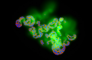
In 2019 Olympus launched TruAI technology, an image analysis approach based on deep neural networks with a focus in segmentation and classification. TruAI technology makes segmentation and classification easy and accessible by non-experts, even in the most difficult scenarios, like in low contrast brightfield images, low signal-to-noise fluorescence images, or in situations of highly dense cell confluency. When installed in the microscope that is acquiring the images, it speeds up the research as soon as analysis results can be available, shortly after the acquisition has concluded, or even at the same time as the acquisition.

In this tech talk, through a collection of examples measured with our live cell imaging systems, high-content screening station, and whole slide scanner, you will see what TruAI technology can do for your research and get a preview of what is coming next.

Presenter: Manoel Veiga, PhD
Application Specialist, Olympus Soft Imaging Solutions

Manoel Veiga earned his PhD in Physical Chemistry in the University Santiago de Compostela, Spain. Following two postdocs in the Universities Complutense of Madrid and WWU Münster, he joined PicoQuant GmbH. After five years supporting customers worldwide in the fields of FLIM and time-resolved spectroscopy, he joined Olympus Soft Imaging Solutions GmbH in 2017, where he works as Global Application Specialist with a focus on high-content analysis and deep learning.


관련 제품

이미징 소프트웨어

cellSens

직관적인 작동 및 원활한 워크플로를 제공하는 cellSens 소프트웨어의 사용자 인터페이스는 맞춤 설정이 가능하기 때문에 레이아웃을 제어할 수 있습니다. cellSens 소프트웨어는 여러 가지 패키지로 제공되며 특정한 이미지화 요구 사항에 최적화된 다양한 기능이 포함되어 있습니다. 그래픽 실험 관리자 및 웰 탐색기 기능은 5D 이미지 획득을 용이하게 합니다. TruSight™ 디콘볼루션을 통해 향상된 해상도를 확보하고 콘퍼런스 모드를 사용하여 이미지를 공유하세요.

  • TruAI™ 딥러닝 분할 분석을 통한 실험 효율 개선으로 라벨 없는 핵 검출 및 세포 계수를 제공하세요.
  • 모듈식 이미지화 소프트웨어 플랫폼
  • 사용자 중심의 인터페이스로 직관적인 애플리케이션
  • 광범위한 기능 세트는 간단한 스냅샷과 고급 다차원 실시간 실험을 지원
Experts
Manoel Veiga
응용 전문가, 생명과학 연구
Olympus Soft Imaging Solutions

안녕하세요. Manoel Veiga입니다. Olympus의 소프트웨어의 딥 러닝을 구현한 팀에 소속되어 있습니다. 2017년에 Olympus에 입사하여 고함량 스크리닝, 이미지 분석 및 딥 러닝의 전문 기술을 개발하였습니다. 형광 수명 이미징의 전문가이기도 합니다. 물리화학 박사 과정 중에 데이터 분석에 처음 관심을 가지게 되었습니다. 이러한 관심은 콘볼루션 신경망의 파워 및 해결할 수 있는 놀라운 이미지 분석 과제를 발견한 후에 더욱 발현되었습니다.

Accelerating Image Analysis with TruAI™ Deep Learning Technology2024년12월12일
죄송합니다. 이 페이지는 해당 국가에서 사용할 수 없습니다.
Ask the Expert Sign-up

By clicking subscribe you are agreeing to our privacy policy which can be found here.

Sorry, this page is not
available in your country.

죄송합니다. 이 페이지는 해당 국가에서 사용할 수 없습니다.