
Spring has officially arrived, and with it comes warmer weather and new life to capture under the microscope. From plants and flowers to fetal cats, here the top images that you thought were the cat’s meow on our Instagram this month.

This image celebrated the first day of spring over on our Instagram account. The star-shaped wildflower known as an Herb Robert (Geranium robertianum) looks beautiful, but it has a surprising secret—when crushed, the petals have an unpleasant odor that repels mosquitoes!
Image courtesy of Karl Gaff.
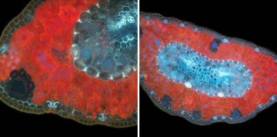
Can you guess what specimen this is? These images show a 20-micron section through a leaf from Pinus sylvestris (commonly known as Scots pine). Native from western Europe to eastern Siberia, the pine is identifiable due to its combination of short, blue-green leaves and orange-red bark. This sample was photographed in UV-induced excitation microscopy.
Image courtesy of Karl Gaff.
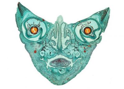
Here we're looking at an amazing cross-section of a fetal cat. Or is the cat looking at us? The microscope image shows maturing ears, eyes, tongue, skull bone, sinus cavities, and other skull features. The sample was 12 mm in diameter.
This stunning image was captured after the fetal cat skull was prepared for examination and cut in a longitudinal view. Four individual images were combined into one final file called a computational photograph, which offers enhanced resolution and image enlargement.
Image courtesy of Mike Peres. Captured using brightfield microscopy at 2x magnification.
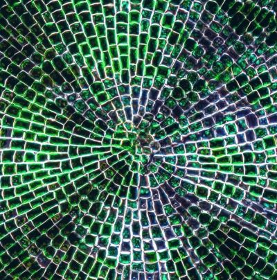
No, this is not mosaic glass or sea glass! What you're looking at is Coleochaete scutata, a flat, green algae that lives attached to other algae, water plants, and flat rocks. This sample was growing on the glass surface of a freshwater aquarium before being collected and photographed under the microscope.
Image courtesy of Håkan Kvarnström.
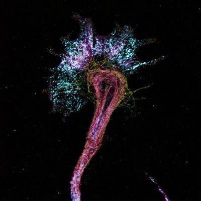
Some people have a lot of nerve! This image reveals the structure of a growth cone on a developing axon terminal. Hippocampal neurons were processed through expansion microscopy (ExM) to magnify nanoscale actin-binding proteins and microtubules components by a massive 10–20-fold increase in size.
To create this image, varying colors were used to reflect the changing depth of the neuronal growth cone as it extended in neural development.
Image courtesy of Arthur Chien. Captured on an Olympus FLUOVIEW FV3000 confocal microscope.
To see more images like these, be sure to follow us on Instagram at @olympuslifescience!
Interested in sharing your own images? Visit our image submission site.
Related Content
From A to Z: Our Most Popular Microscope Images for February 2021
From Algae to Art: Creating Stunning Microscope Artwork with X Line Optics
The Art and Science of Neurogarden—Meet the 2019 IOTY Global Winner
