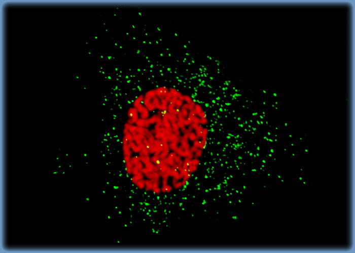
Distribution of Histones and Peroxisomes in Mink Endometrium Epithelial Cells
A log phase culture of GMMe cells was treated with a cocktail of mouse anti-histones (pan) and rabbit anti-PMP 70 (peroxisomal membrane protein) primary antibodies, followed by goat anti-mouse and anti-rabbit secondary antibodies conjugated to Texas Red and Alexa Fluor 488, respectively. The procedure enabled the visualization of the nuclear histone proteins (red emission) and cytoplasmic peroxisomes (green emission) present in the cells.
Sorry, this page is not
available in your country.