The name of J.S. Ploem is almost synonymous with the use of the vertical illuminator for reflected light fluorescence microscopy. Ploem, Brumberg and others were closely associated with the development of dichromatic beamsplitters (dichromatic mirrors) which overcame the light loss problems inherent in the use of ordinary half-mirrors in reflected light microscopy.
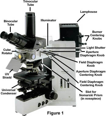
Reflected light fluorescence microscopy is overwhelmingly the choice of today's fluorescence workers. This mode of fluorescence microscopy is also known as incident light fluorescence, epi-fluorescence, or episcopic fluorescence. The universal reflected light vertical illuminator is interposed between the observation viewing tubes and the nosepiece carrying the objectives, as illustrated with the Olympus fluorescence microscope in Figure 1.
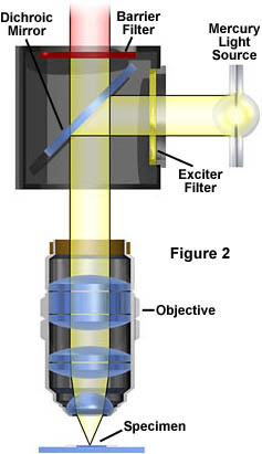
The illuminator is designed to direct light onto the specimen by first passing the light through the microscope objective on the way toward the specimen and then using that same objective to capture the light being emitted by the specimen (diagrammed in Figure 2).
This type of illuminator has several advantages: the objective, first serving as a well corrected condenser and then as the image-forming light gatherer, is always in correct alignment relative to each of these functions; most of the unwanted or unused excitation light reaching the specimen travels away from the objective (such "front-face" fluorescence excitation is particularly good with thick specimens); the area being illuminated is restricted to the area being observed; the full numerical aperture of the objective, in Köhler illumination, is utilizable; and, it is possible to combine or alternate reflected light fluorescence with transmitted light phase contrast, Nomarski differential interference contrast (DIC) or Hoffman modulation contrast observation.
The universal reflected light illuminator (see Figure 1) has at its far end a universal lamp-house which contains a light source, usually a mercury burner (other light sources might be a xenon burner or a halogen bulb.) The light travels along the illuminator parallel to the table top and perpendicular to the optical axis of the microscope. The light passes through collector lenses and a variable, centerable aperture diaphragm and then through a variable, centerable field diaphragm. It is incident upon the excitation filter which selects those excitation wavelengths that are wanted to reach the specimen and blocks the wavelengths not wanted to reach the specimen. The selected wavelengths reach the dichromatic beamsplitting mirror. This mirror is a special type of interference filter which efficiently reflects shorter wavelength light and efficiently passes longer wavelength light. The dichromatic beamsplitter (also sometimes called the dichroic mirror) is tilted at a 45 degree angle to the incident excitation illumination and reflects this light at a 90 degree angle directly through the objective and onto the specimen. The fluorescent light emitted by the specimen is gathered by the objective, now serving in its usual image forming function. Because the emitted light consists of longer wavelengths, it is able to pass through the dichromatic mirror.
Reflected Fluorescence Microscopy
Explore the optical pathways and filter cube combinations in reflected fluorescence microscopy.
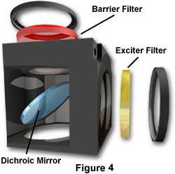
Start Tutorial »Any scattered excitation light reaching the dichromatic mirror is reflected back toward the light source. Before the emitted light can reach the eyepiece, it is incident upon and passes through the barrier or suppression filter. This filter blocks (suppresses) any residual excitation light and passes the desired longer emission wavelengths toward the eyepieces. In most reflected light fluorescence illuminators, the excitation filter, dichromatic mirror, and barrier filter are incorporated in a cube, as illustrated in Figure 4. The more sophisticated systems accommodate three or four fluorescence cubes (on a revolving turret or on a slider) and permit the user to attach replacement custom made exciters, barrier filters or dichromatic mirrors.
The design of the illuminator should permit the user to employ the desirable Köhler Illumination, providing bright and even illumination across the field of view. The corrected condensing lenses of the system make certain that the image of the centerable aperture diaphragm is conjugate with the back aperture of the focused objective. The image of the pre-focused, centerable field diaphragm is conjugate with the focused specimen and the plane of the fixed eyepiece diaphragm.
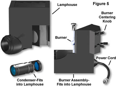
The universal illuminator lamphouse should incorporate an infrared filter to block the very long, heat generating wavelengths. Some lamphouses have a built-in red suppression filter (e.g. an BG38), or a slot for such a filter, to eliminate a reddish background seen in the field of view in some applications. The lamphouse itself should not leak harmful ultraviolet wavelengths and, preferably, should incorporate a switch to automatically shut down the lamp if the housing is inadvertently opened during operation. The lamphouse should be sturdy enough to withstand a possible burner explosion during operation. The lamp socket should have lamp centering screws to permit centering the image of the lamp arc or halogen lamp coil to the back aperture of the objective (in Köhler illumination these planes are conjugate).
An ultraviolet protection shield is fitted into the front of the illuminator to protect the user's eyes from any inadvertent leakage of potentially dangerous short wavelength radiation. In the light path, closer to the lamphouse and before the excitation filter, it is desirable to have a light shutter for complete blocking of excitation light. The light shutter thus permits you to block the burner light without switching the burner off; neutral density filters permit reduced intensity to diminish fading in some specimens.
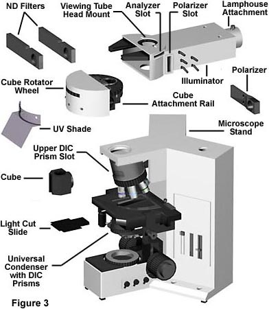
The universal reflected light illuminator can be attached to the standard modular upright microscope; and a similar version is now used with inverted microscope stands. The inverted stands also permit combining or alternating between reflected light fluorescence and the various contrast techniques of transmitted light microscopy.
The vertical illuminator, preferably, should have no magnification factor. Some illuminators have a magnification factor of 1.25X and are so inscribed.