Application of silicone immersion objectives to long-term live-cell imaging of plant zygote embryogenesis
Plant zygote embryogenesis
Studies on embryogenesis in the field of life science research have made progress together with advances in microscopy techniques. In particular, studies on animal embryogenesis have developed into research on stem cells such as embryogenic stem (ES) cells. In contrast, plant embryogenesis is far less understood than animal embryogenesis even though it has been studied for a long time. The major reason for this slow progress is the difficulty of live observation of the division process of the
fertilized egg cell (zygote) because the angiosperm zygote is deeply embedded in the pistil, a maternal tissue.
The Optical Technology Group of ERATO Higashiyama Live-Holonics Project, Nagoya University, a research group led by Dr. Daisuke Kurihara, has been working on elucidating the plant reproductive system by primarily using live-cell imaging and microscopic cell manipulation techniques. Recently, they succeeded in live-cell imaging of the plant zygote embryogenesis process, a world first. This used to be considered to be very difficult. The results from a series of
studies were published in Developmental Cell*, an American scientific journal, in July 2015. In this study, long-term live-cell imaging of plant zygote division and growth was made possible by development of a special medium and a new microdevice.
This application note introduces an example of long-term live-cell imaging of plant zygote embryogenesis using a silicone immersion objective, which is specially designed for live-cell imaging and was used in this study.
Application of silicone immersion objectives
Long-term live-cell imaging of Arabidopsis zygote embryogenesis
1)Establishment of a culture method for long-term real-time observation of embryogenesis in live ovules
Angiosperm embryogenesis occurs within the ovule, which is deeply embedded in the pistil (Fig. 1); therefore, it has been impossible to observe embryogenesis from zygote division in living material.
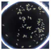
The Optical Technology Group of ERATO Higashiyama Live-Holonics Project, Nagoya University, a research group led by Dr. Daisuke Kurihara, used the model plant Arabidopsis thaliana, removed ovules from the pistils, and examined the composition of a medium that would enable embryogenesis in an ovule under in vitro culture conditions. By examining the medium composition that supports normal embryo development and ovule growth, a significant increase in the frequency
of ovule survival and normal embryo development was observed when trehalose, an organic substance, was added to the culture medium for the ovary, a pistil tissue surrounding the ovule. Development of this new ovule culture medium allowed development of the embryo into a mature seed within the ovule outside the pistil, germination, and growth of the seedling into an adult plant (Fig. 2).
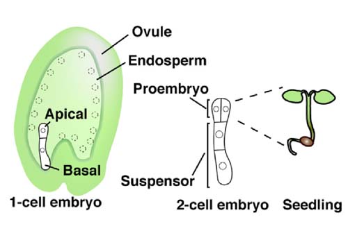
Fig1. Schematic representation of an Arabidopsis flower and embryogenesis
After significant elongation of the zygote in the ovule, it divides asymmetrically into a smaller apical cell and a larger basal cell. The apical cell generates the embryo, which ultimately gives rise to the adult plant.
 Day7 | 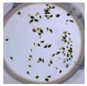 Day10 | 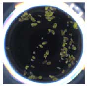 Day14 | 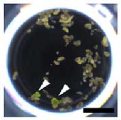 Day19 |
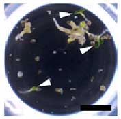 Day26 | 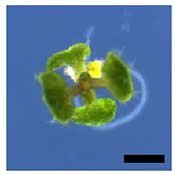 Day32 | 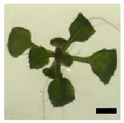 Day37 |
Fig2. Growth of early embryos to seedlings in ovule culture media, and a seedling into an adult plant
Next, the group of Dr. Kurihara worked on development of a microdevice that enabled stable long-term imaging. Since the ovule is oval shaped, it moves during observation and may wander outside the microscopic field. Because of this, stable live observation of the process of embryogenesis occurring over several days was not possible. By improving their proprietary microcage array that can stably hold ovules, they developed a micropillar array. This is a new microdevice that can stably hold ovules for a long period without preventing their growth. A new system for stable long-term culture of the ovule in a fixed position was established by development of this new microdevice and the ovule culture medium.
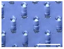 Tilted-view | 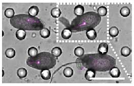 Top-view | 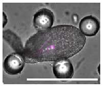 |
Fig3. Fixation of ovules with the microdevice
A micropillar array of a uniform pillar distribution is used for stable long-term retention of ovules. In-depth observation of embryogenesis is possible because pillars hold ovules without preventing their growth. Scale bars represent 300 mm.
2)Use of silicone immersion objectives for long-term live-cell imaging of Arabidopsis zygote embryogenesis
The world’s first long-term and real-time observation of embryogenesis a zygote to late embryo was made possible by combining the ovule culture system described above and a microscope system capable of high-sensitivity imaging of the embryo, which is inside the ovule covered by multiple layers of cells.
The high-sensitivity microscope system used in this study was the fully-motorized research inverted microscope IX series with a 30´ silicone immersion objective, UPLSAPO30XS.
The silicone immersion objectives are specially designed for live-cell imaging with an optical design based on the refractive index of silicone oil (ne≈1.40), which is close to that of living tissue (ne≈1.38). The 30´ silicone immersion objective, UPLSAPO30XS, used in this imaging experiment has a high numerical aperture of 1.05 and long working distance of 0.8 mm, which is required for deep, high-definition imaging while retaining a wide field.
Unlike water immersion objectives which require a water supply because of evaporation, it is not necessary to supply silicone oil because it does dry out at 37°C during operation over several days. In addition, the silicone immersion objectives are compatible with the Z-Drift Compensation System IX-ZDC of the fully-motorized research inverted microscope IX series. Live images that are constantly in focus can be obtained by using a silicone immersion objective with the Z-Drift Compensation
System IX-ZDC.
As an example of live-cell imaging using a 30´ silicone immersion objective, UPLSAPO30XS, the process of Arabidopsis embryogenesis was stably observed in real time for 67 hours from the early embryo (4-cell stage) to the late embryo (Movie).
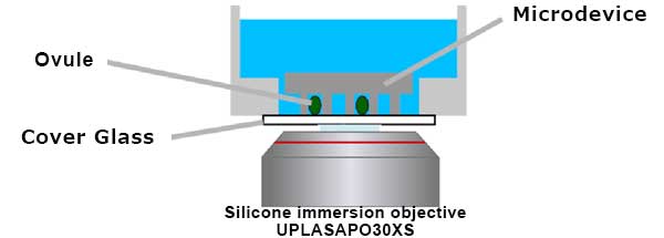
Fig4. Schematic illustration of microdevice imaging with a silicone immersion objective
Multi-point time-lapse imaging was acquired using a motorized XY stage
Movie. Live-cell imaging of zygote division and embryogenesis
The process of embryogenesis from the early embryo (4-cell stage) to the late embryo was recorded over 67 hours by taking images at 10-minute intervals. The movie captured formation of a round-shaped tissue from proembryo cells while changing the orientation of divisions, and formation of rod-like tissue from suspensor cells, which divide only transversely. The numbers indicate the time from the beginning of recording. Nucleus and plasma membrane are labeled green (H2B-GFP) and magenta
(tdTomato-LTI6b), respectively.
Imaging conditions
Imaging system: Fully motorized research inverted microscope IX series
Objective: silicone immersion objective UPLSAPO30XS
Confocal scanner unit: CSU-X1 (Yokogawa Electric Corporation)
EMCCD camera: Evolve 512 (Photometrics)
Motorized XY stage: MD-XY30100T-Meta (Molecular Devices)
Piezo Z-focus drive: P-721 (Physik Instrumente)
In parallel with this study using a live-cell imaging technology, Dr. Kurihara’ research group also developed the ClearSee method, which is a new technique for optical clearing of a whole plant, and published their results in Development, an American scientific journal, in October 2015.
Global application of the newly developed live-cell imaging technology and the new optical clearing technique for plants is expected to bring about major progress in plant research.
This application note was prepared with the help of
Dr. Daisuke Kurihara, Group Leader, Optical Technology Group, ERATO Higashiyama Live-Holonics Project, Nagoya University
For more details on the studies in this application note, please refer to the articles below.
*Source: Dev Cell. 2015 Jul 27;34(2):242-51. doi: 10.1016/j.devcel.2015.06.008. Epub 2015 Jul 9.
Journal: Developmental Cell
Publication date: July 9, 2015
Title: Live-Cell Imaging and Optical Manipulation of Arabidopsis Early Embryogenesis
Authors: Keita Gooh, Minako Ueda, Kana Aruga, Jongho Park, Hideyuki Arata, Tetsuya Higashiyama, and Daisuke Kurihara
**Source: Development. 2015 Dec 1;142(23):4168-79. doi: 10.1242/dev.127613. Epub 2015 Oct 22.
Journal: Development
Publication date: October 22, 2015
Title: ClearSee: a rapid optical clearing reagent for whole-plant fluorescence imaging
Authors: Daisuke Kurihara, Yoko Mizuta, Yoshikatsu Sato, and Tetsuya Higashiyama
Silicone immersion objectives for realization of deep, high-definition 3D live-cell imaging over a prolonged period
Olympus has a line-up of 30/40/60/100´ silicone immersion objectives (UPLSAPO30XS/UPLSAPO40XS/UPLSAPO60XS2/UPLSAPO100XS), which offer both a high numerical aperture and long working distance. Since the refractive index of silicone oil (ne≈1.40) is close to that of living tissue (ne≈1.38), spherical aberration induced by a refractive-index mismatch is reduced in the observation of thick living tissues, thereby enabling high-resolution imaging. In addition, silicone oil does not require the extra task of supplying immersion liquid because it does not dry out or becomes solid. The silicone immersion objectives are compatible with the Z-Drift Compensation System IX-ZDC of the fully-motorized research inverted microscope IX series. Long-term, stable high-resolution 3D imaging that is constantly in focus is achieved and is unaffected by temperature changes or the addition of reagent solution.
Produtos usados nesta aplicação
Sorry, this page is not
available in your country.