Nome: descrição
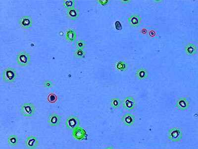
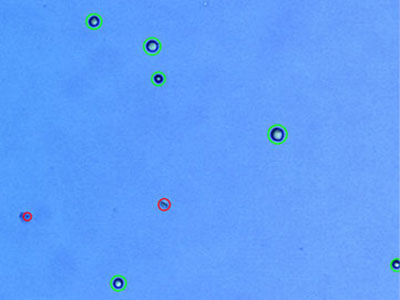
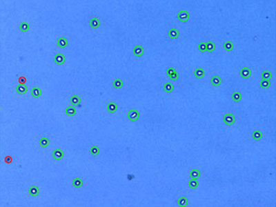
HiF FF: Células iPS humanas sem alimentador (linha celular 201B7)
 | |
Protocolo
|
Resultados
|
|---|
Redução de ruído: 6
Circularidade: 60%
Tamanho mínimo da célula (μm): 3
Tamanho máximo da célula (μm): 60
Nível de desagrupamento: médio
|
Concentração total de células (células/ml): 1,50 × 10⁶
Concentração de células vivas (células/ml): 1,38 × 10⁶
Concentração de células mortas (células/ml): 1,19 × 10⁵
Viabilidade (%): 92,0
Tamanho médio da célula (μm): 15,9
|
|
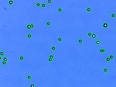
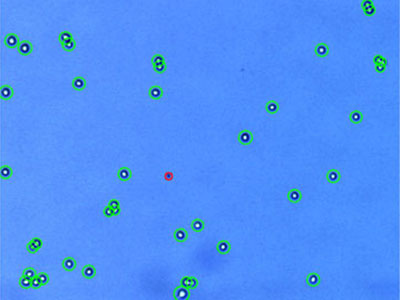
MEF: Fibroblastos embrionários de murino
 | |
Protocolo
|
Resultados
|
|---|
Redução de ruído: 7
Circularidade: 60%
Tamanho mínimo da célula (μm): 3
Tamanho máximo da célula (μm): 60
Nível de desagrupamento: nenhum
|
Concentração total de células (células/ml): 1,03 × 10⁶
Concentração de células vivas (células/ml): 9,37 × 10⁵
Concentração de células mortas (células/ml): 9,72 × 10⁴
Viabilidade (%): 90,6
Tamanho médio da célula (μm): 19,6
|
|
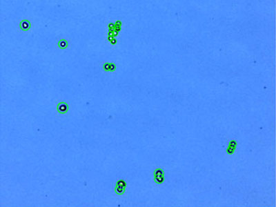
A549:Carcinoma pulmonar humano
 | |
Protocolo
|
Resultados
|
|---|
Redução de ruído: 5
Circularidade: 60%
Tamanho mínimo da célula (μm): 3
Tamanho máximo da célula (μm): 60
Nível de desagrupamento: médio
|
Concentração total de células (células/ml): 1,09 × 10⁶
Concentração de células vivas (células/ml): 1,07 × 10⁶
Concentração de células mortas (células/ml): 1,78 × 10⁴
Viabilidade (%): 98,4
Tamanho médio da célula (μm): 17,5
|
|
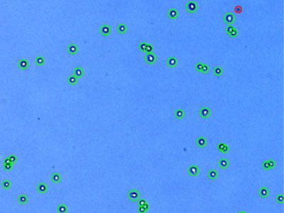
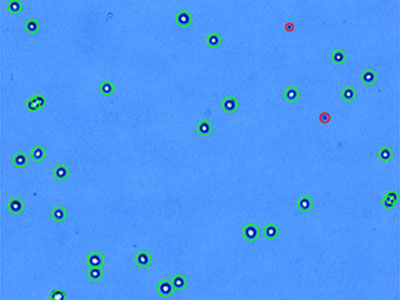
AsPC-1: Adenocarcinoma pancreático humano
 | |
Protocolo
|
Resultados
|
|---|
Redução de ruído: 5
Circularidade: 60%
Tamanho mínimo da célula (μm): 3
Tamanho máximo da célula (μm): 60
Nível de desagrupamento: alto
|
Concentração total de células (células/ml): 7,51 × 10⁵
Concentração de células vivas (células/ml): 7,19 × 10⁵
Concentração de células mortas (células/ml): 3,17 × 10⁴
Viabilidade (%): 95,8
Tamanho médio da célula (μm): 10,7
|
|
C6: Glioma de rato
 | |
Protocolo
|
Resultados
|
|---|
Redução de ruído: 5
Circularidade: 60%
Tamanho mínimo da célula (μm): 3
Tamanho máximo da célula (μm): 60
Nível de desagrupamento: médio
|
Concentração total de células (células/ml): 1,31 × 10⁶
Concentração de células vivas (células/ml): 1,22 × 10⁶
Concentração de células mortas (células/ml): 8,91 × 10⁴
Viabilidade (%): 93,2
Tamanho médio da célula (μm): 12,1
|
|
Capan-2: Adenocarcinoma pancreático humano
 | |
Protocolo
|
Resultados
|
|---|
Redução de ruído: 5
Circularidade: 60%
Tamanho mínimo da célula (μm): 3
Tamanho máximo da célula (μm): 60
Nível de desagrupamento: médio
|
Concentração total de células (células/ml): 1,04 × 10⁶
Concentração de células vivas (células/ml): 1,00 × 10⁶
Concentração de células mortas (células/ml): 3,61 × 10⁴
Viabilidade (%): 96,5
Tamanho médio da célula (μm): 13,4
|
|
DU 145: Carcinoma da próstata humana
 | |
Protocolo
|
Resultados
|
|---|
Redução de ruído: 5
Circularidade: 60%
Tamanho mínimo da célula (μm): 3
Tamanho máximo da célula (μm): 60
Nível de desagrupamento: médio
|
Concentração total de células (células/ml): 1,86 × 10⁶
Concentração de células vivas (células/ml): 1,80 × 10⁶
Concentração de células mortas (células/ml): 5,34 × 10⁴
Viabilidade (%): 97,1
Tamanho médio da célula (μm): 14,3
|
|
293-GFP: HEK293 expressando GFP de forma estável
 | |
Protocolo
|
Resultados
|
|---|
Redução de ruído: 5
Circularidade: 60%
Tamanho mínimo da célula (μm): 3
Tamanho máximo da célula (μm): 60
Nível de desagrupamento: médio
|
Concentração total de células (células/ml): 1,84 × 10⁶
Concentração de células vivas (células/ml): 1,82 × 10⁶
Concentração de células mortas (células/ml): 2,23 × 10⁴
Viabilidade (%): 98,8
Tamanho médio da célula (μm): 11,3
|
|
H1299: Carcinoma de pulmão de células não pequenas humanas
 | |
Protocolo
|
Resultados
|
|---|
Redução de ruído: 5
Circularidade: 60%
Tamanho mínimo da célula (μm): 3
Tamanho máximo da célula (μm): 60
Nível de desagrupamento: médio
|
Concentração total de células (células/ml): 4,14 × 10⁵
Concentração de células vivas (células/ml): 3,52 × 10⁵
Concentração de células mortas (células/ml): 6,23 × 10⁴
Viabilidade (%): 84,9
Tamanho médio da célula (μm): 19,5
|
|
H4: Neuroglioma humano
 | |
Protocolo
|
Resultados
|
|---|
Redução de ruído: 5
Circularidade: 60%
Tamanho mínimo da célula (μm): 3
Tamanho máximo da célula (μm): 60
Nível de desagrupamento: médio
|
Concentração total de células (células/ml): 2,08 × 10⁵
Concentração de células vivas (células/ml): 1,44 × 10⁵
Concentração de células mortas (células/ml): 6,32 × 10⁴
Viabilidade (%): 69,6
Tamanho médio da célula (μm): 12,8
|
|
HCT116: Carcinoma colorretal humano
 | |
Protocolo
|
Resultados
|
|---|
Redução de ruído: 5
Circularidade: 60%
Tamanho mínimo da célula (μm): 3
Tamanho máximo da célula (μm): 60
Nível de desagrupamento: médio
|
Concentração total de células (células/ml): 1,34 × 10⁶
Concentração de células vivas (células/ml): 1,32 × 10⁶
Concentração de células mortas (células/ml): 1,81 × 10⁴
Viabilidade (%): 98,7
Tamanho médio da célula (μm): 12,2
|
|
HeLa:Carcinoma cervical humano*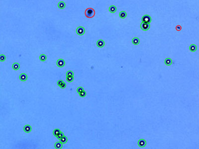 | |
Protocolo
|
Resultados
|
|---|
Redução de ruído: 5
Circularidade: 60%
Tamanho mínimo da célula (μm): 3
Tamanho máximo da célula (μm): 60
Nível de desagrupamento: médio
|
Concentração total de células (células/ml): 8,98 × 10⁵
Concentração de células vivas (células/ml): 8,11 × 10⁵
Concentração de células mortas (células/ml): 8,69 × 10⁴
Viabilidade (%): 90,3
Tamanho médio da célula (μm): 17,6
|
|
Hep3B: Carcinoma hepatocelular humano
 | |
Protocolo
|
Resultados
|
|---|
Redução de ruído: 5
Circularidade: 60%
Tamanho mínimo da célula (μm): 3
Tamanho máximo da célula (μm): 60
Nível de desagrupamento: médio
|
Concentração total de células (células/ml): 1,54 × 10⁶
Concentração de células vivas (células/ml): 1,49 × 10⁶
Concentração de células mortas (células/ml): 5,87 × 10⁴
Viabilidade (%): 96,2
Tamanho médio da célula (μm): 14,9
|
|
HL-60: Leucemia promielocítica aguda humana
 | |
Protocolo
|
Resultados
|
|---|
Redução de ruído: 5
Circularidade: 60%
Tamanho mínimo da célula (μm): 3
Tamanho máximo da célula (μm): 60
Nível de desagrupamento: médio
|
Concentração total de células (células/ml): 1,57 × 10⁶
Concentração de células vivas (células/ml): 1,45 × 10⁶
Concentração de células mortas (células/ml): 1,23 × 10⁵
Viabilidade (%): 92,2
Tamanho médio da célula (μm): 14,9
|
|
Hs 578T: carcinoma mamário humano
 | |
Protocolo
|
Resultados
|
|---|
Redução de ruído: 5
Circularidade: 60%
Tamanho mínimo da célula (μm): 3
Tamanho máximo da célula (μm): 60
Nível de desagrupamento: médio
|
Concentração total de células (células/ml): 1,13 × 10⁶
Concentração de células vivas (células/ml): 8,72 × 10⁵
Concentração de células mortas (células/ml): 2,62 × 10⁵
Viabilidade (%): 76,9
Tamanho médio da célula (μm): 16,5
|
|
MCF7:Adenocarcinoma mamário humano
 | |
Protocolo
|
Resultados
|
|---|
Redução de ruído: 5
Circularidade: 60%
Tamanho mínimo da célula (μm): 3
Tamanho máximo da célula (μm): 60
Nível de desagrupamento: médio
|
Concentração total de células (células/ml): 4,74 × 10⁵
Concentração de células vivas (células/ml): 4,43 × 10⁵
Concentração de células mortas (células/ml): 4,07 × 10⁴
Viabilidade (%): 91,4
Tamanho médio da célula (μm): 16,2
|
|
NIH3T3: Fibroblasto embrionário de camundongo
 | |
Protocolo
|
Resultados
|
|---|
Redução de ruído: 5
Circularidade: 60%
Tamanho mínimo da célula (μm): 3
Tamanho máximo da célula (μm): 60
Nível de desagrupamento: médio
|
Concentração total de células (células/ml): 1,52 × 10⁶
Concentração de células vivas (células/ml): 1,33 × 10⁶
Concentração de células mortas (células/ml): 1,96 × 10⁵
Viabilidade (%): 87,1
Tamanho médio da célula (μm): 14,1
|
|
PANC-1:Carcinoma ductal pancreático humano
 | |
Protocolo
|
Resultados
|
|---|
Redução de ruído: 5
Circularidade: 60%
Tamanho mínimo da célula (μm): 3
Tamanho máximo da célula (μm): 60
Nível de desagrupamento: alto
|
Concentração total de células (células/ml): 1,23 × 10⁶
Concentração de células vivas (células/ml): 1,21 × 10⁵
Concentração de células mortas (células/ml): 2,26 × 10⁴
Viabilidade (%): 98,2
Tamanho médio da célula (μm): 15,3
|
|
PC-3: Adenocarcinoma da próstata humano
 | |
Protocolo
|
Resultados
|
|---|
Redução de ruído: 5
Circularidade: 60%
Tamanho mínimo da célula (μm): 3
Tamanho máximo da célula (μm): 60
Nível de desagrupamento: médio
|
Concentração total de células (células/ml): 1,36 × 10⁶
Concentração de células vivas (células/ml): 1,33 × 10⁶
Concentração de células mortas (células/ml): 3,16 × 10⁴
Viabilidade (%): 97,7
Tamanho médio da célula (μm): 17,2
|
|
STO: Fibroblasto embrionário em rato
 | |
Protocolo
|
Resultados
|
|---|
Redução de ruído: 5
Circularidade: 0%
Tamanho mínimo da célula (μm): 3
Tamanho máximo da célula (μm): 60
Nível de desagrupamento: médio
|
Concentração total de células (células/ml): 1,78 × 10⁶
Concentração de células vivas (células/ml): 1,50 × 10⁶
Concentração de células mortas (células/ml): 2,81 × 10⁵
Viabilidade (%): 84,2
Tamanho médio da célula (μm): 13,1
|
|
U-2 OS: Osteossarcoma humano
 | |
Protocolo
|
Resultados
|
|---|
Redução de ruído: 5
Circularidade: 60%
Tamanho mínimo da célula (μm): 3
Tamanho máximo da célula (μm): 60
Nível de desagrupamento: médio
|
Concentração total de células (células/ml): 7,66 × 10⁵
Concentração de células vivas (células/ml): 7,21 × 10⁵
Concentração de células mortas (células/ml): 4,45 × 10⁴
Viabilidade (%): 94,2
Tamanho médio da célula (μm): 16,0
|
|
U937: Linfoma humano
 | |
Protocolo
|
Resultados
|
|---|
Redução de ruído: 5
Circularidade: 40%
Tamanho mínimo da célula (μm): 3
Tamanho máximo da célula (μm): 60
Nível de desagrupamento: médio
|
Concentração total de células (células/ml): 1,22 × 10⁶
Concentração de células vivas (células/ml): 1,12 × 10⁶
Concentração de células mortas (células/ml): 1,02 × 10⁵
Viabilidade (%): 91,6
Tamanho médio da célula (μm): 13,4
|
|
* Embora tenha se tornado uma das linhagens celulares mais importantes na pesquisa médica, é importante reconhecer que a contribuição de Henrietta Lacks para a ciência aconteceu sem o seu consentimento. Essa injustiça, embora tenha levado a descobertas importantes em imunologia, doenças infecciosas e câncer, também levantou discussões importantes sobre privacidade, ética e consentimento na medicina.
Para saber mais sobre a vida de Henrietta Lacks e sua contribuição para a medicina moderna, clique aqui.
http://henriettalacksfoundation.org/
|