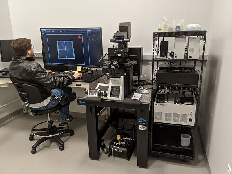
Dynamic Volumetric Imaging with the FV3000RS Confocal Microscope: 3D Reconstruction of Actin Dynamics in a Colletotrichum graminicola Spore
Colletotrichum graminicola is a widespread fungal pathogen and a major disease-causing agent of cereal crops, including wheat and corn. In corn, fungal infections of C. graminicola cause anthracnose leaf blight, resulting in severe crop damage and major economic losses for farmers. Understanding the cytoskeletal dynamics in germinating C. graminicola spores could therefore contribute to the prevention of disease.
Pregermination Organization of Filamentous Actin Cables
In this experiment, the FLUOVIEW FV3000RS microscope was used to study the actin dynamics of a C. graminicola spore prior to germination. Filamentous actin cables can be observed within a crescent-shaped spore that has yet to germinate. The cables have organized to extend lengthwise across the spore at the plane of contact with the surface. The actin cables can be seen retracting from the center towards either end of the spore.
Figure 1. XYZT reconstruction of actin dynamics in a spore of C. graminicola before germination.
Imaging conditions
Objective: 100X oil 1.49NA TIRF objective (UAPON100XOTIRF)
Microscope: FLUOVIEW FV3000RS laser scanning confocal microscope
Lasers: 488 nm (GFP, green), 561 nm (DIC, gray)
Scanner: Resonant scanner
XY frames: 0.272 sec/frame
Z series: 22 steps (6 sec/section)
T series: 30 Z stacks acquired over 3 minutes
Observation of Two Distinct Cytoskeletal Events during Germination
Following the organization of actin into cables, the germination process of a C. graminicola spore is marked by two successive cytoskeletal events, with the formation of both patches and cables during both events. In the first event, the cytoskeleton rearranges to form a septum that delineates the fungal spore into two distinct cells (depicted by the medial barrier across the width of the spore). Following the septation event, actin cables form at the site of germination and extend into the developing germ tube toward the apex. These two events can be seen in Figures 2 and 3.
Figure 2. XYZT of cross-wall formation during a septation event in a germinating spore of C. graminicola, followed by the formation of a germ tube.
Imaging conditions
Objective: 100X oil 1.49NA TIRF objective (UAPON100XOTIRF)
Microscope: FLUOVIEW FV3000RS laser scanning confocal microscope
Lasers: 488 nm (GFP, green), 561 nm (DIC, gray)
Scanner: Resonant scanner
XY frames: 0.470 sec/frame
Z series: 31 steps (5.35 sec/stack)
T series: 34 Z stacks acquired in 1-minute intervals over 33 minutes
Figure 3. 3-D reconstruction of the actin cytoskeleton in a germinating spore of C. graminicola, in which the developing germ tube can clearly be seen.
Imaging conditions
Objective: 100X oil 1.49NA TIRF objective (UAPON100XOTIRF)
Microscope: FLUOVIEW FV3000RS laser scanning confocal microscope
Lasers: 488 nm (GFP, green), 561 nm (DIC, gray)
Scanner: Galvano scanner
Z sections: 17 steps
How the FV3000 Confocal Microscope Facilitated Our Experiment
Maintaining Focus during 4-Dimensional Time-Lapses with Olympus’ TruFocus Z-Drift Compensation System
The advanced TruFocus Z-drift compensation system (ZDC) facilitates the acquisition of multiple sequential Z stacks with no drift in the Z dimension. This enables users to generate clear, focused, and continuous time lapses in three dimensions, which enables researchers to interrogate protein dynamics in the entire sample without concern for Z-drift.
Rapid Four-Dimensional Acquisition with the Resonant Scanner
The FV3000RS microscope features a high-performance resonant scanner that enables rapid acquisition time-lapses of live-cell dynamics. The resonant scanner in the FV3000RS microscope also maintains a full 1X field of view with FN 18, with imaging speeds of 30 frames per second at 512 × 512 pixels. Using the resonant scanner, researchers can acquire Z stacks of numerous planes in seconds, achieving high-resolution Z stacks of rapidly occurring phenomena in live cells.
TruSpectral GaAsP Detectors Provide High Sensitivity for Live Cell Imaging
Integrated in all FV3000 confocal microscopes, TruSpectral detection technology enables higher light throughput compared to conventional spectral detection units. The volume phase hologram diffracts light with an up to three-fold higher transmission efficiency than reflection gratings. When combined with high-efficiency GaAsP PMTs, the FV3000 microscope’s high-sensitivity spectral detector (HSD) requires minimal laser power to obtain high signal-to-noise images, enabling powerful yet gentle live cell imaging.
Comment by Joseph Vasselli and Dr. Brian Shaw
Our research has the goal of understanding the role of the cytoskeleton and endocytic proteins in growth and development in filamentous fungi. Tracking the spatiotemporal localization of these proteins during shifts in growth polarity is crucial to our experiments. The FV3000 microscope’s superb Z-drift compensation, high-sensitivity detectors, and rapid resonant scanner enables us to acquire high-resolution XYZTs to effectively interrogate the dynamics of these proteins in live cells.

Acknowledgments
This application note was prepared with the help of the following researchers:
| Dr. Brian D. Shaw, Professor, Fungal Biology, Department of Plant Pathology and Microbiology, Texas A&M University |
| Joseph Vasselli, Graduate Student, Department of Plant Pathology and Microbiology, Texas A&M University |
适于这类应用的产品
Maximum Compare Limit of 5 Items
Please adjust your selection to be no more than 5 items to compare at once
对不起,此内容在您的国家不适用。

