Eingesetztes Produkt

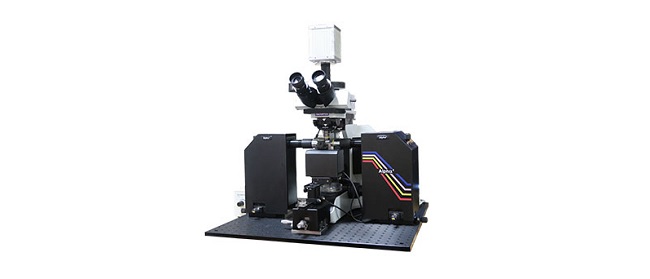
Alpha3
Das moderne Fluoreszenz-Lichtscheibenmikroskop bietet eine flexible, hochleistungsfähige Lichtscheibenmikroskopielösung für die Forschung. Unter Verwendung einer intelligenten optischen Fokusverschiebung wird eine gleichmäßige, artefaktfreie Bildgebung über das gesamte Sichtfeld ermöglicht. Das Alpha3 System kann je nach den spezifischen Anforderungen leicht mit den Optionen, wie Smart 3D Scanning, XY-Kacheln und Temperaturregelung, aufgerüstet werden.
- Schnelle, artefaktfreie 3D-Bildgebung dank multidirektionaler Lichtscheibe mit Echtzeit-Fokusabtastung
- Kompatibel mit einer großen Auswahl an hochwertigen Olympus Optiken
- Zubehör für eine Vielzahl von Proben mit Beständigkeit gegen organische Lösungsmittel oder wässrige Puffer
- Benutzerfreundliche QtSPIM Software und Workstation für nahtlose 3D-Bilderfassung bei maximaler Geschwindigkeit
.jpg?rev=4058)
CM20
Remotely monitor, analyze, and share your cell cultures’ health, cell count, and confluency using the reliable quantitative data provided by the automated CM20 incubation monitoring system. The system enables label-free observation, reduces the risk of damage to your cultures, and standardizes your culture workflow.
- Automatically collects quantitative data on the health and confluency of your cultures
- Monitor, analyze, and share your cultures' progress remotely from a PC or tablet
- Equipped with oblique epi-illumination for label-free observation

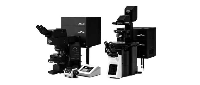
FV3000
- Erhältlich als reine Galvanometer-Konfiguration (FV3000) oder als Hybrid-Galvanometer-/Resonanzscanner-Konfiguration (FV3000RS)
- Neue hocheffiziente und genaue TruSpectral Erkennung auf allen Kanälen
- Optimiert für die Bildgebung von lebenden Zellen mit hoher Empfindlichkeit und geringer Fototoxizität
- Inverted and upright frame options to suit a variety of applications and sample types

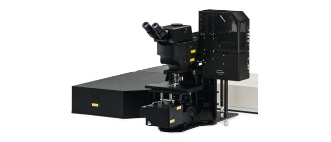
FVMPE-RS
- Maximale Auflösung und maximaler Kontrast mit TruResolution-Objektiven
- High-Speed-Bildgebung mit Resonanz-Scanner
- Erweiterte IR-Multiphotonenanregung bis 1300 nm
- Optionaler Dreifachscanner für Multiphotonen- und Laserstimulation im Bereich des sichtbaren Lichts
- Hocheffiziente Transmission mit optischen Beschichtungen bis 1600 nm auf speziellen Multiphotonenobjektiven und Scanning-Einheit
- Automatische Ausrichtung der IR-Laserstrahlen in 4 Achsen

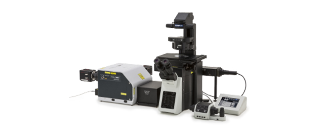
IXplore Spin (Inactive)
- Rapid and high-resolution confocal imaging with a spinning disk system
- 3D confocal time-lapse imaging of live cells with less phototoxicity and bleaching
- Precise 3D imaging with improved light collection using silicone oil immersion objectives
- Upgrade to the IXplore SpinSR super resolution system depending on your research progress and/or budget
.jpg?rev=8603)
NoviSight
Die 3D-Zellanalyse-Software NoviSight liefert statistische Daten für Sphäroide und 3D-Objekte in Mikrotiterplatten-Experimenten. Damit lassen sich die Zellaktivität in 3D quantifizieren und die Nachweisempfindlichkeit verbessern, seltene Zellereignisse können leichter erfasst und Zellzahlen genauer bestimmt werden. Die NoviSight Software ist für verschiedene Bildgebungsverfahren geeignet, d. h. von der konfokalen Point-Scan-Bildgebung, der Zwei-Photonen-Bildgebung und der konfokalen Spinning-Disk-Bildgebung bis hin zur hochauflösenden Lebendzell-Bildgebung.
- Schnelle 3D-Bilderkennung von ganzen Strukturen bis hin zu subzellulären Merkmalen
- Genaue statistische Analyse
- Ausgestattet mit einer Vielzahl einsatzbereiter Standard-Assays oder einfache Erstellung eigener Assays
