The objective is perhaps the most important part of an optical microscope because it’s responsible for primary image formation and plays a central role in determining image quality. But with so many microscope objective types available, it can be difficult to find the right one for your application. Where do you start? In today’s blog post, we compiled a series of questions to help you weigh your options and choose the right objective for your application. Let’s dive in. |  |
1. What is the size of your specimen?
Olympus’ microscope objectives feature a range of magnifications from 1.25x to 150x. This is the first parameter to consider when finding the best objective for your application. Combined with the magnification from the eyepieces, it determines the microscope’s overall magnification.
2. What are the smallest features in your specimen that you want to observe?
The second most important parameter of a microscope objective is the numerical aperture (NA). NA measures its ability to gather light. It’s an important factor to determine resolution, depth of focus, and the brightness of images. Objectives with a larger NA gather a wider range of light, resulting in brighter, higher resolution images.
NA is also important to observe very fine structures or detect dim signals during fluorescence observation. When determining which microscope objective will resolve the smallest feature in your specimen, think about the NA. As you weigh your options, keep in mind that numerical aperture ranges between 0.04 to 1.7.
3. What are your image field-of-view and depth-of-field requirements?
Two important microscope objective characteristics are field number and depth of field.
- The field-of-view number, or field number, is the diameter of the field of view in your optical microscope. It is expressed in millimeters and measured at the intermediate image plane. Keep in mind, modern plan apochromats and other specialized flat-field objectives often feature a usable field that ranges between 22 and 26.5 millimeters (or more when combined with widefield eyepieces).
- The objective depth of field is the axial range, which enables you to focus an objective without any considerable change in image sharpness. This value varies radically from low to high numerical aperture objectives; it usually decreases as the numerical aperture increases.
4. What is your resolution requirement?
The resolution of the microscope objective determines the smallest distance between two objects that can be observed. It is directly proportional to the illumination wavelength of light and inversely proportional to the NA.
The higher the NA, the smaller the distance between two objects. As we mentioned previously, choosing the right NA for your application is crucial in determining the resolution of your microscope system.
5. What working distance do you need?
Working distance (WD) is the distance from the objective’s front lens to the closest surface of the coverslip when the specimen is in focus. WD is inversely proportional to the NA, which means that higher NA objectives typically have low working distances.
If your application demands non-coverslip applications that require an increased distance between the focal plane and the objective’s tip (i.e. samples with irregular topography, delicate structures, or mechanical constraints of the overall optical assembly), then you may find Olympus’ LMPLFLN or SLMPLN long-working distance objectives well-suited to your application.
6. If you have a fluorescence sample, how bright is your fluorescence signal?
If you work with samples that emit a weak fluorescence signal, then we recommend high NA objectives since they gather more light. Olympus has a wide range of microscope objectives that offer fluorescence excitation from ultraviolet (UV) to near-infrared (NIR).
7. Are you using multichannel fluorescence imaging or one channel?
To compensate for chromatic correction, you can use different types of objectives: achromat, semi-apochromat, and apochromat. Achromat objectives provide the least amount of correction, semi-apochromats (or fluorites) have additional spherical corrections, and apochromats offer the highest chromatic correction.
If your application demands multichannel fluorescence, then we recommend Olympus UPLXAPO extended apochromat objectives.
8. What is your observation method?
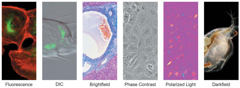
When dealing with samples that are too thick, too thin, or birefringent, researchers will apply different observation methods other than brightfield. Common techniques are darkfield, differential interference contrast (DIC), phase contrast, and polarization. Olympus offers dedicated objectives associated with these different observation methods.
9. What is the medium your sample is in?
Many microscope objectives are designed to image specimens with air as the medium, while others use an immersion medium that contains a higher refractive index to enable a high NA and resolution.
For instance, using immersion oil instead of air as the imaging medium can increase the resolution by a factor of approximately 1.5. The most common immersion media are air, water, oil, and silicone. Choosing the appropriate objective for your media will result in better image quality. | 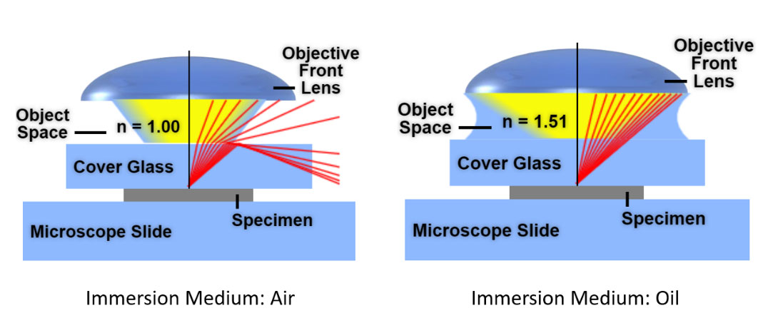 |
10. Are you using an advanced microscope system?
Olympus offers dedicated objectives for advanced optical systems such as confocal, spinning disk confocal, multi-photon excitation, and total internal reflection fluorescence (TIRF) microscopy. If you work with these systems, then choosing the correct objective for your application is crucial.
Additional resources and tools to find the right objective
Choosing the right objective can improve your microscope’s imaging performance and produce more reliable results for quantification and analysis. To help you make this important decision, we compiled some helpful resources:
- Use Olympus’ objective finder to select your application requirements, compare objectives, and determine which objective best suits your application.
- Learn more about microscope objective specifications and identification.
If you have any questions about our objectives, don’t hesitate to reach out.
Related Content
Quick Guide for Choosing the Right MPE Objectives
Video: Silicone Oil Immersion Objectives for Live Cell Imaging
White Paper: X Line Objectives Enable Reliable Data Acquisition Over a Large Field of View