Evident LogoOlympus Logo

Ask the Experts

Olympus Bioimaging Conference: Exploring New Dimensions | 3-Day Virtual Event | March 9–11, 2022

Bio Imaging Conference

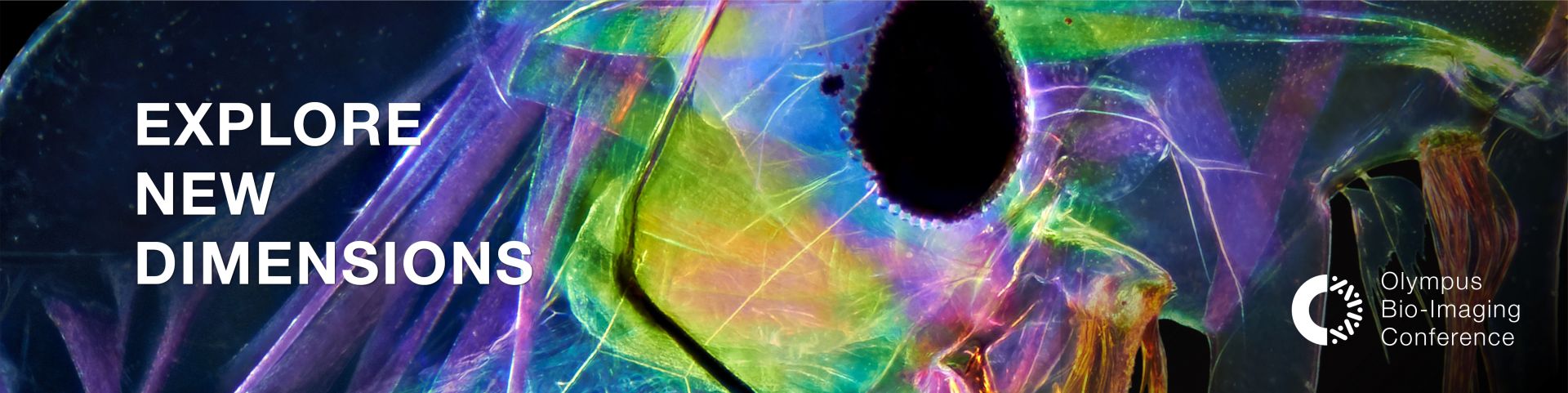
Bioimaging is an essential tool used to image and analyze cells, tissues, and molecules in fields such as drug discovery, diagnostics, life science, and clinical research. Rapid advances in imaging technology have enabled researchers to visualize and quantify specimens in ways that were, until recently, impossible. With the advent of technologies such as super-resolution and AI-assisted imaging, researchers can now clearly observe their specimens on the nanometer scale and have access to stress-free, accurate data analysis. The objective of this virtual conference is to explore and understand recent innovations in light microscopy bioimaging technologies. Over the 3-day event, our expert speakers will discuss topics such as super-resolution microscopy, F-techniques including FRAP and FRET, and novel probes.

Register Now


Agenda

Time (GMT +8) Day 1
March 9, 2022
Day 2
March 10, 2022
Day 3
March 11, 2022

1:30 p.m.–1:40 p.m.

Welcome Address

Presenter: Mr. Kefeng Wang, Director of Life Science Sales, Scientific Solutions Business Division, Olympus China

Chairperson Address

Presenter: Dr. Qian Peter Su, Principal Investigator, University of Technology Sydney (UTS)

Welcome Address

Presenter: Mr. Sam Habib, Head of APAC Sales, Scientific Solutions Business Division, Olympus Corporation of Asia Pacific

Chairperson Address

Presenter: Prof. Sarah Ellis, Head of the Centre for Imaging the Tumour Environment (CITE), Australia

Welcome Address

Presenter: Mr. Olivier Dupuis, Head of APAC Marketing, Scientific Solutions Business Division, Olympus Corporation of Asia Pacific

Chairperson Address

Presenter: Dr. Graham Wright, Director, Research Support Centre (RSC) A*STAR, Singapore

1:40 p.m.–2:25 p.m.

Overcoming Physical Resolution Limits of Fluorescence Microscopes with Sparse Deconvolution

Speaker: Dr. Liangyi Chen, College of Future Technology, Peking University

More information

Applications of Fluorescence Resonance Energy Transfer and Fluorescence Recovery After Photobleaching In Vivo Using Drosophila as a Model System

Speaker: Dr. Krishanu Ray, Department of Biological Science, Tata Institute of Fundamental Research, India

More information

Gentle Probes for 4D Imaging

Speaker: Dr. Zhixing Chen, College of Future Technology, Peking University

More information

2:25 p.m.–3:10 p.m.

Upconversion Nanophotonic Systems for Super-Resolution Imaging, Single Molecular Tracking, and High-Throughput Digital Assays

Speaker: Prof. Dayong Jin, University of Technology Sydney and Southern University of Science and Technology, Australia

More information

Nanoscale Biophotonics: Using Multimodal Imaging to Understand the Inner Workings of the Body

Speaker: Prof. Brant Gibson, RMIT University, Melbourne, Australia

More information

Complex Regulation of Mitochondrial Calcium Uniporter Complex

Speaker: Dr. Karthik Mallilankaraman, Yong Loo Lin School of Medicine, National University of Singapore

More information

3:10 p.m.–3:45 p.m.

Stunning Details in Living Cells Using Olympus’ IXplore SpinSR System

Speaker: Shaoling Qi, Olympus China

More information

Arabidopsis Autophagy-Related 3 (ATG3) Facilitates the Liquid–Liquid Phase Separation of ATG8e to Promote Autophagy

Speaker: Dr. Bin Guan, School of Agriculture and Biology, Shanghai Jiao Tong University

More information

Mitochondrial Relocation of a Common Synthetic Antibiotic: A Nongenotoxic Approach to Cancer Therapy

Speaker: Prof. Jong Seung Kim, Department of Chemistry, Korea University

More information

3:45 p.m.–4:30 p.m.

Higher, Faster, Stronger: Fluorescent Imaging for Cytoskeleton-Related Activities

Speaker: Dr. Xueliang Zhu, Shanghai Institute of Biochemistry and Cell Biology, Chinese Academy of Sciences

More information

Live Demo: Fast, Accurate, and Flexible Photomanipulation Using the Olympus IXplore™ Spin System and cellFRAP Module

Demonstrator: Mitsuru Araki, Olympus China

More information

Live Demo: Olympus FLUOVIEW™ FV3000 Near-Infrared Confocal Laser Scanning Microscope

Demonstrators: Srivats Hariharan & Guo Lin, Olympus Singapore

More information


Speakers

Dr. Liangyi Chen

College of Future Technology
Peking University

Dr. Krishanu Ray

Department of Biological Science
Tata Institute of Fundamental Research, India

Dr. Liangyi Chen

Biography

Dr. Liangyi Chen is Boya Professor of Peking University. He obtained his undergraduate degree in biomedical engineering from Xi’an JiaoTong University, then majored in biomedical engineering in pursuing a PhD at Huazhong University of Science and Technology. His lab focused on two interweaved aspects: the development of new imaging and quantitative image analysis algorithms and the application of these technologies to study how glucose-stimulated insulin secretion is regulated in the health and disease at multiple levels (single cells, islets, and in vivo) in the health and disease animal models. The techniques developed included ultrasensitive Hessian structured illumination microscopy (Hessian SIM) for live cell super-resolution imaging, the sparse deconvolution algorithm for extending spatial resolution of fluorescence microscopes limited by the optics, super-resolution fluorescence-assisted diffraction computational tomography (SR-FACT) for revealing the three-dimensional landscape of the cellular organelle interactome, two-photon three-axis digital scanned light sheet microscopy (2P3A-DSLM) for tissue and small organism imaging, and fast high-resolution miniature two-photon microscopy (FHIRM-TPM) for brain imaging in freely behaving mice. He is also recipient of the National Distinguished Scholar Fund project from the National Natural Science Foundation of China.

Abstract

Overcoming Physical Resolution Limits of Fluorescence Microscopes with Sparse Deconvolution

During this session, Dr. Chen will present two novel high-resolution fluorescence microscopy methods his lab invented for live sample imaging:

"The first one is for live-cell long-term super-resolution (SR) imaging. We developed a deconvolution algorithm for structured illumination microscopy based on Hessian matrixes (Hessian-SIM). It uses the continuity of biological structures in multiple dimensions as a priori knowledge to guide image reconstruction and attains artifact-minimized SR images with less than 10% of the photon dose used by conventional SIM while substantially outperforming current algorithms at low signal intensities. Its high sensitivity allows the use of sub-millisecond excitation pulses followed by dark recovery times to reduce photobleaching of fluorescent proteins, enabling hour-long time-lapse SR imaging in live cells.

After this initial work, we realized that the spatial resolutions of live-cell super-resolution microscopes are limited by the maximum collected photon flux. Taking advantage of a priori knowledge of the sparsity and continuity of biological structures, we develop a deconvolution algorithm that further extends the resolution of super-resolution microscopes under the same photon budgets by nearly twofold. As a result, sparse structured illumination microscopy (Sparse-SIM) achieves ~60 nm resolution at a 564 Hz frame rate, allowing it to resolve intricate structural intermediates, including small vesicular fusion pores, ring-shaped nuclear pores formed by different nucleoporins, and relative movements between the inner and outer membranes of mitochondria in live cells. Likewise, sparse deconvolution can be used to increase the three-dimensional resolution and contrast of spinning-disc confocal-based SIM (SD-SIM) and operates under conditions with the insufficient signal-to-noise ratio, all of which allows routine four-color, three-dimensional, ~90 nm resolution live-cell super-resolution imaging. Overall, we argue that sparse deconvolution may be a general tool to push the spatiotemporal resolution limits of live-cell fluorescence microscopy."

Dr. Krishanu Ray

Biography

Krishanu Ray is a Fellow of the Indian National Science Academy and a Professor at the Department of Biological Sciences, Tata Institute of Fundamental Research (TIFR), Mumbai, India. He completed his Master’s in Biophysics, Molecular Biology and Genetics from the University of Calcutta and PhD in Molecular Biology from TIFR under the aegis of Mumbai University. He then worked at the Institute of Molecular Cell Biology, Singapore and Howard Hughes Medical Institute, University of California, San Diego, before joining TIFR. He established the laboratory at TIFR in 1998 to investigate the molecular cell biology of motor proteins and study their impact on the development and behavior of an organism. His research focuses on how kinesin-2, a molecular motor, moves soluble and vesicle-associated proteins in axons and cilia in response to external stimuli. He also studies how strain induces contractile actomyosin assembly at the cell membrane. He utilizes microscopic tools to visualize protein-protein interactions and dynamics, as well as the subcellular distribution of fluorescent proteins in Drosophila neurons and other tissues.

Abstract

Applications of Fluorescence Resonance Energy Transfer and Fluorescence Recovery after Photobleaching In Vivo Using Drosophila as a Model System

Fluorescence spectroscopy is utilized to monitor a variety of molecular states and parameters. The introduction of confocal microscopy and genetically coded fluorescent proteins expanded the scope of applying this technique in situ to cellular and subcellular scales for monitoring functional properties and distribution of endogenous proteins in their natural milieu. This seminar will discuss applications of two specific methods—fluorescence resonance energy transfer (FRET) and fluorescence recovery after photobleaching (FRAP)—in live Drosophila tissues. FRET is a molecular-scale ruler that can determine distances between two parts of a protein and between two proteins. It can be appraised from the fluorescent life-time data and changes in the acceptor and donor emissions. We used this technique to assess the interaction dynamics between the kinesin-2 motor subunits and between choline acetyltransferase and kinesin-2 motor in axons. FRAP is a simple and powerful tool to determine the flow of molecules inside a cell. We monitored the movement of soluble proteins such as the choline acetyltransferase in the axon, odorant receptor co-receptor in the cilia and F-actin dynamics in somatic cells surrounding mature spermatids using FRAP. Applications of these tools provided critical insights into the states of the protein-protein interactions and dynamics within a cell in specific biological contexts.

Dr. Zhixing Chen

College of Future Technology
Peking University

Prof. Dayong Jin

University of Technology Sydney and Southern University of Science and Technology, Australia

Dr. Zhixing Chen

Biography

Dr. Zhixing Chen received a BSc in chemical biology from Tsinghua University (2008) and a PhD in chemistry from Columbia University (2014, with Profs. Virginia Cornish and Wei Min). He had additional training at Stanford University (Postdoc 2016–2018 with Prof. Yan Xia), Columbia University (Postdoc 2015), and Peking University (RA 2008–2009). Zhixing is a chemist with research experience spanning natural product synthesis, polymer chemistry, fluorescence and non-linear optical probes, bioconjugation chemistry, and live cell imaging. His notable achievements include research involving a vibrational palette, an isotopically edited alkyne and nitrile group for multiplexed Raman imaging, and ladderane unzipping, using mechanical force to reconfigure polymer from an insulating material to a semiconductor. Zhixing’s current focus is to develop new imaging tools with high biocompatibility to promote advanced bioimaging technologies.

Abstract

Gentle Probes for 4D Imaging

Modern fluorescence-imaging methods promise to unveil organelle dynamics in live cells. Phototoxicity, however, has become a prevailing issue when boosted illumination applies. From a chemistry perspective, Dr. Chen presents how to engineer organic dyes with minimal phototoxicity with two examples on mitochondrial markers and insulin secretion markers. Resonating with the ongoing theme of reducing photodamage using optical approaches, these biocompatible probes promise to offer additional spatial-temporal information in the era of 4D physiology.

Prof. Dayong Jin

Biography

Dr. Dayong Jin is a Distinguished Professor at the University of Technology Sydney (UTS) since 2017 and a Chair Professor at Southern University of Science and Technology since 2019.

Professor Jin obtained his PhD from Macquarie University in 2007. At Macquarie, he was promoted to Lecturer in 2010, Senior Lecturer in 2013, Associate Professor in 2014, and Professor in 2015.

At UTS, as the director, he established the Australian Industry Transformation Research Hub for Integrated Devices for End-user Analysis at Low Levels (ARC IDEAL Hub), the Department of Industry, Science, Energy and Resources’ Australia-China Joint Research Centre for Point of Care Testing (DISER POCT), the UTS-SUStech Joint Research Centre for Biomedical Materials & Devices, which three major programs underpin the UTS Institute for Biomedical Materials & Devices (IBMD), to transform advances in phonics and materials into disruptive biotechnologies.

His research has been in the physical, engineering, and interdisciplinary sciences, with expertise covering biomedical optics, nanotechnology, microscopy, diagnostics, and microfluidics devices.

Prof. Jin is the winner of the Australian Museum Eureka Prize for Interdisciplinary Scientific Research (2015), the Australian Academy of Science John Booker Medalist (2017), and the Prime Minister’s Prize for Physical Scientist of the Year 2017. In 2021, Professor Jin won the Australian Laureate Fellowship and was elected to the fellowship of Australian Academy of Technology and Engineering.

Abstract

Upconversion Nanophotonic Systems for Super-Resolution Imaging, Single Molecular Tracking, and High-Throughput Digital Assays

Dr. Jin will showcase the recent advances made in nanophotonics, biophonics, and quantumbiophotonics that enable their transformation into cellular probes and sensors with single-molecule sensitivity.

“In parallel to the developments of molecular probes, we have invented new modalities and devised a series of new instruments to acquire high-dimensional and super-resolution details of living cells.

In my talk, I will present the recent advances in a new family of nanophotonic “Super Dots” that can up-convert infrared photons into intense visible light at the nanoscale. Each single Super Dot can be highly doped with more than 10^4 lanthanide ions for high brightness and nonlinear optical responses. Several fascinating properties have been discovered since to allow high-throughput bio-discoveries, data storage, single nanoparticle lasing and high-security-level anti-counterfeiting applications, setting records for the tracking of single-molecule transport, super-resolution microscopy, nanoscale thermometry, and recently super-capacity single molecule digital assays and optical tweezers. Our research will enable super-resolution imaging of single molecules and live cells in their physiological environment, to watch subcellular compartments at work and understand the nanoscale world inside living cells. This will provide a novel toolkit, analogous to Google’s ‘street view’, that will enable researchers to ‘drop down’ and observe the details of subcellular ‘live traffic’, decode the complexities of life science machinery and detect health issues before they become critical."

Prof. Brant Gibson

RMIT University
Melbourne, Australia

Dr. Karthik Mallilankaraman

Yong Loo Lin School of Medicine
National University of Singapore

Prof. Brant Gibson

Biography

Professor Gibson was awarded his PhD from La Trobe University in 2004. From 2005-09, he was a photonics development engineer at Quantum Communications Victoria (QCV) where he and colleagues designed and developed Australia’s first commercial quantum security product (QCV SPS 1.01). In 2011, he was awarded an Australian Research Council (ARC) Future Fellowship on Hybrid Diamond Materials for Next Generation Sensing, Biodiagnostic and Quantum Devices. Prof. Gibson is currently in the joint roles of Assistant Associate Dean (Physics, RMIT University), Deputy Director, and RMIT Node Leader of the ARC Centre of Excellence for Nanoscale BioPhotonics and Deputy Director of the Sir Lawrence Wackett Centre for Defence at RMIT University. He has wide-ranging research interests in the areas of diamond, fluorescent nanoprobes, wide band gap materials, single photon sources, quantum sensors, hybrid nanomaterial integration, fiber optics, photonics, biophotonics, optical, confocal, and atomic force microscopy, and he has published more than 100 refereed journal publications.

Abstract

Nanoscale Biophotonics: Using Multimodal Imaging to Understand the Inner Workings of the Body

Light-based imaging and sensing tools can assist with our understanding of the complex chemical and molecular processes taking place in and around cells in the living body [1]. Fluorescent nanodiamonds (NDs) are an attractive nanoscale-tool that have a range of unique properties which make them highly desirable for bioimaging and biosensing applications [2]. Their fluorescence is produced via optical excitation of atomic defects, such as the negatively charged nitrogen vacancy centre, within the diamond crystal lattice. Possessing long-wavelength emission, high brightness, no photobleaching, no photoblinking, nanometer size, a room temperature sensitivity to magnetic and microwave fields, and an exceptional resistance to chemical degradation make NDs almost the ideal fluorescent bioimaging nanoprobe [3]. I will discuss these exciting properties in detail and give some examples of the effect of surface functionality on their fluorescent properties [4], their use as fluorescent probes for pH [5] and hydrogen peroxide sensing in biological systems [6], and the effect of particle size on nanodiamond fluorescence and colloidal properties in biological media [7]. In addition, I will also discuss hybrid biosensing applications including the incorporation of NDs into polycaprolactone [8] and silk [9] electrospun materials.

Dr. Karthik Mallilankaraman

Biography

Dr. Karthik Mallilankaraman obtained his PhD in Medical Microbiology from the University of Madras, India. He moved to the University of Pennsylvania in Philadelphia, Pennsylvania to complete a post-doctoral fellowship with Professor David Weiner on developing DNA vaccines to the Chikungunya virus. He then moved to Temple University in Philadelphia to study the mitochondrial calcium uniporter, where he identified the regulatory subunit of the uniporter MCUR1 and discovered the gatekeeper of the uniporter, which establishes the setpoint of mitochondrial calcium uptake. For these contributions he received 2013 Young Bioenergeticist Award from Biophysical Society. To further understand the mitochondrial matrix calcium regulation, he moved back to the University of Pennsylvania (Foskett lab) and worked on regulatory mechanisms of the mitochondrial calcium uniporter. Dr. Karthik moved to Singapore in 2015 and started his independent research group at the Department of Physiology in YLL School of Medicine, National University of Singapore.

Abstract

Complex Regulation of Mitochondrial Calcium Uniporter Complex

Cellular respiration in eukaryotes occur in both aerobic and anaerobic modes; however, a majority of the energy is derived from aerobic respiration. The aerobic phases occur within organelles called mitochondria in two steps: the Krebs Cycle and the electron transport chain. Mitochondria are sometimes referred to as the “power plants” of the cell, as these are the organelles that generate most of the cell’s supply of adenosine triphosphate (ATP). Interestingly, the aerobic respiration is regulated by an important second messenger calcium. Mitochondrial matrix calcium acts as a cofactor in both Krebs cycle and electron transport chain.

My talk will focus on how calcium enters the mitochondrial matrix through a tightly regulated ion channel embedded in the inner mitochondrial membrane. Mitochondrial calcium uniporter (MCU) is the Ca2+-selective ion channel localized in the inner mitochondrial membrane (IMM) that mediates Ca2+ uptake into the mitochondrial matrix from the cytoplasm to regulate metabolism, cell death, and cytoplasmic Ca2+ signaling. The mitochondrial Ca2+ uniporter is a complex of proteins including the pore-forming subunit MCU and accessory proteins including MICU1, MICU2, MCUR1, and EMRE. We discovered MCUR1 as the positive regulator of the uniporter as well as the role of MICU1 in gatekeeping the MCU. Loss of MCUR1 ablates mitochondrial calcium uptake and blunts ATP production activating AMPK mediated pro-survival autophagy. But loss of MICU1 results in constitutive MCU activation, enhanced mitochondrial Ca2+ uptake at low cytoplasmic [Ca2+] and mitochondrial Ca2+ overload. We were the first to show MICU1 provided a gatekeeping function that reduces the permeability of the uniporter in situ below a threshold value of 1-3 μM external free Ca2+ to prevent mitochondrial Ca2+ overload under basal conditions. While controversy existed on the localization of MICU1, a majority of studies, including our recent work, suggests inter-membrane space localization of MICU1 and MICU2. Thus, mitochondria are protected both from Ca2+ depletion under low-Ca2+ conditions and from Ca2+ overload under normal resting conditions by a unique molecular complex that involves Ca2+ sensors on both sides of the IMM, MICU1, and MICU2 from the IMS side and the unknown matrix Ca2+ sensing components of the uniporter complex.

Shaoling Qi

Olympus China

Dr. Bin Guan

School of Agriculture and Biology
Shanghai Jiao Tong University

Shaoling Qi

Biography

Shaoling Qi is senior product manager for life science at Olympus China. She joined Olympus Life Science as application specialist right after receiving master’s degree from School of Life Science, Tsinghua University in 2009. With years of hands-on experience and application knowledge of advance microscopy, Shaoling helps scientists identify and apply imaging solutions that support and advance their research goals. She is currently in charge of the customization business in China and responsible for life science product management, marketing, and sales support of high-end imaging systems, such as confocal, multiphoton, and super resolution technology.

Abstract

Stunning Details in Living Cells Using Olympus’ IXplore SpinSR System

Live cell super-resolution imaging is now a growing application trend in life science, clinical research, and regenerative medicine studies. However, there is always a trade-off between spatial resolution, temporal resolution, and phototoxicity, making it a significant challenge to investigate fine structural dynamics in cells. We have developed both hardware and software solutions to overcome these challenges. In this session, I will discuss how Olympus’ IXplore SpinSR system, combines speed, sensitivity, and resolution for live-cell-compatible super resolution imaging. I will also be presenting several application examples where the Olympus IXplore SpinSR system has been effective.

Dr. Bin Guan

Biography

Dr. Bin Guan is currently a postdoctoral fellow in the School of Agriculture and Biology, Shanghai Jiao Tong University. Bin Guan received his Ph.D. degree in Genetics from National Key Laboratory of Plant Molecular Genetics, CAS Center for Excellence in Molecular Plant Sciences, Chinese Academy of Sciences (CAS) in 2021. His research interest includes phospholipids and autophagy.

Abstract

Mitochondrial Relocation of a Common Synthetic Antibiotic: A Nongenotoxic Approach to Cancer Therapy

Phase separation plays an important role in various physiological and signalling processes in plant and animal cells by forming relatively independent spatial domains that selectively enrich molecules and form unique structures. Autophagy, a highly regulated degradation mechanism in eukaryotic cells, has been shown to degrade liquid condensates, and pre-autophagosomal structures (PAS) also undergo liquid-liquid phase separation to regulate autophagosome formation. Although the ubiquitin-like protein ATG8 plays a central role in modifying autophagosomes and recruiting specific cargoes to autophagosomes, it is unclear whether ATG8 undergoes phase separation to regulate autophagosome biosynthesis. In this study, systematic cytological observations revealed that Arabidopsis ATG8e can phase separate in vivo and in vitro, and that intrinsically disordered regions (IDR) at its N-terminal are involved in phase separation formation. Treatment with phase separation inhibitors and observation of genetic material suggest that liquid-liquid phase separation of ATG8e plays an important role in autophagy. Further studies have shown that the autophagy-associated protein ATG3 enhances the phase separation of ATG8e and in turn promotes autophagy. Interestingly, the N-terminal region of ATG8e's homologues in yeast and mammals, Atg8 and LC3 (three members), is also IDR, suggesting that phase separation may exist between mammalian LC3 and yeast Atg8 and that phase separation is involved in the regulation of autophagy in mammalian cells. This study demonstrates the existence and importance of phase separation in autophagy, and provides important clues for the in-depth study of the molecular regulation mechanism of autophagy.

Prof. Jong Seung Kim

Department of Chemistry
Korea University

Dr. Xueliang Zhu

Shanghai Institute of Biochemistry and Cell Biology
Chinese Academy of Sciences

Prof. Jong Seung Kim

Biography

Jong Seung Kim received his PhD from the Department of Chemistry and Biochemistry at Texas Tech University in 1993. He also has one year of post-doctoral research experience at the University of Houston. Currently, he is a full professor in the Department of Chemistry at Korea University in Seoul. He has been a member of the Korean Academy of Science and Technology since 2014. He has published around 530 papers in prestigious journals with an h-index of 104. His research interests are application of organic chemistry in drug delivery and theranostics of various pathologies, including Alzheimer’s disease and malignant neoplasms and their super-resolution imaging. He has been named as a Highly Cited Researcher since 2014.

Abstract

Mitochondrial Relocation of a Common Synthetic Antibiotic: A Nongenotoxic Approach to Cancer Therapy

Tumor recurrence because of therapy-induced nuclear DNA lesions is a major issue in cancer treatment. Currently, only a few examples of potentially nongenotoxic drugs have been reported. Dr. Kim discusses here a new approach to generating such leads that involves the mitochondrial re-localization of ciprofloxacin, one of the most prescribed synthetic antibiotics. A rationally designed conjugation strategy, involving the linking of ciprofloxacin to a triphenyl phosphonium group (giving lead Mt-CFX), is used to enhance the concentration of ciprofloxacin in the mitochondria of cancer cells. Support for this proposed localization came from an analogue of Mt-CFX bearing a fluorescent probe. Localization of Mt-CFX to the mitochondria induces oxidative damage to proteins, mtDNA, and lipids. A large bias in favor of mtDNA damage over nDNA was seen with Mt-CFX, whereas the opposite result was obtained for several classic cancer chemotherapeutics. Mt-CFX was found to produce a statistically significant reduction in cancer growth in a xenograft mouse model. It also proved to be generally well tolerated. Taken in concert, these findings led Dr. Kim and his colleagues to suggest mitochondrial relocalization of antibiotics could emerge as a useful approach to generating anticancer leads that promote cell death via the selective induction of mitochondrially-mediated oxidative damage.

Dr. Xueliang Zhu

Biography

Dr. Xueliang Zhu received his BS (1985) and MS (1988) in Department of Biology, University of Science and Technology of China and was employed as an assistant lecturer. He went to University of California, San Diego and the Institute of Biotechnology, University of Texas Health Science Center at San Antonio for his dissertation as a joint graduate student (1990-1994) and received his PhD (1995) from the Shanghai Institute of Cell Biology, Chinese Academy of Sciences (CAS). After his post-doctoral training (1995-1997) at the Shanghai Research Center of Life Sciences, CAS, he became a PI at the center. He moved to the Shanghai Institute of Biochemistry and Cell Biology in 1999. He is a cell biologist, mainly interested in cytoskeleton-dependent subcellular structures and functions.

Abstract

Higher, Faster, Stronger: Fluorescent Imaging for Cytoskeleton-Related Activities

Spatiotemporal resolution is the major limitation factor in imaging subcellular structures or their dynamic activities. Cilia, for instance, are approximately 250-nm in diameter but contain elegant ultrastructure and bidirectional intraflagellar transport (IFT) machineries critical for protein import from and export into the cell body. Furthermore, motile cilia can beat swiftly (10 Hz or more) and exist in hundreds per cell. In this presentation, Dr. Zhu shares some of his and his colleague’s imaging experiences achieved during their research that rival even the latest cutting-edge fluorescent microscopes.

Mitsuru Araki

Olympus China

Srivats Hariharan

Olympus Singapore

Mitsuru Araki

Biography

Mr. Mitsuru Araki is a Deputy General Manager of the Sales Support Department in Olympus China. He holds a bachelor’s degree in Applied Physics from Tohoku University, Japan. He joined Olympus Japan in 2009 as a technical service engineer. In 2012, he transferred to Olympus India and established a technical service team for the microscopy business. In 2015, he moved back to Japan to accept the position of product manager for imaging software and sales support for the Chinese life science market. In 2020, he transferred to Olympus China and is currently in charge of life science product marketing and sales support.

Abstract

Live Demo: Fast, Accurate, and Flexible Photomanipulation Using the Olympus IXplore™ Spin System and cellFRAP Module

Photomanipulation is an essential factor for various imaging techniques, such as FRAP, FLIP, photoactivation, uncaging, etc., to study protein dynamics in living cells. For example, in FRAP, a specific area of a fluorescent dye or a fluorescence-labelled protein in a cell is bleached, and the recovery of fluorescence is observed to examine fluidity of the target. These imaging techniques are employed in various application fields; however, accurately stimulating the target in a dynamically moving living cell is still challenging. Olympus’ IXplore Spin system offers fast confocal imaging and our cellFRAP module makes accurate, fast, and flexible photostimulation possible. In this session, we will demonstrate the benefit of the IXplore Spin and cellFRAP combination for FRAP experiments.

Srivats Hariharan

Biography

Mr. Srivats Hariharan is an Applications & Marketing Manager for the Olympus Asia-Pacific (APAC) region. He holds a bachelor’s degree in Mechanical Engineering from Nanyang Technological University, Singapore. He has experience working in biomedical research labs and an A*STAR Microscopy Core Facility where he supported researchers on confocal and live cell imaging technologies and helped set up single-molecule super-resolution and light sheet microscopes. He joined the life science team of Olympus Singapore in 2011 as a Product Manager and is in charge of supporting research customers and business partners in South-East Asia and Taiwan. He is currently in charge of all life science marketing related activities in APAC.

Abstract

Live Demo: Olympus FLUOVIEW™ FV3000 Near-Infrared Confocal Laser Scanning Microscope

Two-photon excitation microscopes, such as the Olympus FVMPE-RS system, have long been seen as an ideal choice for deep imaging as they use infrared lasers that scatter less and significantly reduce photobleaching and phototoxicity in fixed and live biological samples. However, there has been a growing demand for single-photon systems, such as the Olympus FV3000 laser scanning confocal microscope, that are capable of using near-infrared (NIR) for fluorescence multiplexing, deep imaging, and live cell imaging. Combined with Olympus’s advanced X Line and A Line objective lenses, NIR imaging can provide clear and high-resolution images deep within the biological specimen. Organic dyes and fluorescent proteins inlucing Cy7, Cy7.5, Alexa Fluor750, Alexa Fluor790, and iRFPs can now be easily imaged along with regular markers in the visible range. In this session, we will demonstrate how to set up and use the Olympus FV3000 microscope for NIR imaging.

Guo Lin

Olympus Singapore

Guo Lin

Biography

Dr. Lin obtained his PhD in 2010 from the National University of Singapore, working on biophysical research. In 2009, he joined Olympus as a Technical and Application Specialist for our laser-based high-end imaging systems. In 2012, Lin decided to move back to China, taking a position with one of the leading scientific camera manufacturers, Photometrics. There, he started as an application specialist, later became a regional sales manager, and finally a scientific sales manager for the Asia-Pacific region. In 2021, Lin moved back to Singapore, joining Olympus Singapore as a products and applications manager. Lin has extensive experience with diverse scientific digital imaging techniques, including various camera technologies.

Abstract

Live Demo: Olympus FLUOVIEW™ FV3000 Near-Infrared Confocal Laser Scanning Microscope

Two-photon excitation microscopes, such as the Olympus FVMPE-RS system, have long been seen as an ideal choice for deep imaging as they use infrared lasers that scatter less and significantly reduce photobleaching and phototoxicity in fixed and live biological samples. However, there has been a growing demand for single-photon systems, such as the Olympus FV3000 laser scanning confocal microscope, that are capable of using near-infrared (NIR) for fluorescence multiplexing, deep imaging, and live cell imaging. Combined with Olympus’s advanced X Line and A Line objective lenses, NIR imaging can provide clear and high-resolution images deep within the biological specimen. Organic dyes and fluorescent proteins inlucing Cy7, Cy7.5, Alexa Fluor750, Alexa Fluor790, and iRFPs can now be easily imaged along with regular markers in the visible range. In this session, we will demonstrate how to set up and use the Olympus FV3000 microscope for NIR imaging.

Olympus Bioimaging Conference: Exploring New Dimensions | 3-Day Virtual Event | March 9–11, 2022Apr 18 2024
Sorry, this page is not available in your country
Ask the Expert Sign-up

By clicking subscribe you are agreeing to our privacy policy which can be found here.

Sorry, this page is not
available in your country.

Sorry, this page is not available in your country