Just a few years ago, Laurent Formery’s image of a sea urchin received an honorable mention in our Image of the Year 2020 competition. Now his stunning image of a sea star has won the global award in our 2022 edition, making him the star of our light microscopy imaging contest and earning him the grand prize—a brand new SZX7 stereo microscope with a DP23 digital camera.
In this interview, Laurent tells us more about his winning image, approach to the art of science, and view on marine life through the microscope lens.
Q: What does your winning image show?
Laurent: This image shows the nervous system of a sea star (Patiria miniata). The main feature of the nervous system in sea stars are three long tracts of neurons called radial nerve cords, which run on the oral side along the midline of each of the five arms. These five nerve cords are linked together by a nerve ring that circles around the mouth. From the nerve cords, regularly spaced lateral nerves branch toward the edges of each arm.
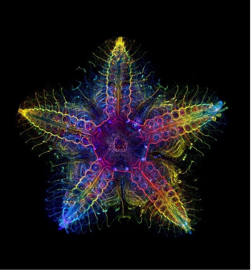
Captured by Laurent Formery (USA), this image of a nervous system of a juvenile sea star (Patiria miniata) won the global award in our Image of the Year 2022 competition.
Q: What do you find exciting about the image and subject matter?
Laurent: As surprising as it seems, sea stars are closely related to vertebrates (the group of animals to which we belong). Yet, they couldn’t look more different than us at first glance. Evolution has gone wild in the lineage of the tree of life containing sea stars and their kind (sea urchins, sea cucumbers), giving these animals a fivefold symmetry completely different from us and other related animals. Because they are so different, we know very little about them: how their nervous system is wired, how it works, and how it compares with ours.
Q: How did you create the image?
Laurent: This image shows a whole-mount immunostaining using an anti-acetylated-tubulin antibody, which is a marker of the nervous system. The sample is very large (about 1 cm) and was imaged at 5X magnification with tile-scanning on a confocal microscope. Stacks of pictures were taken along the oral-aboral axis of the animal, and a depth color-coded Z-projection was done using Fiji software. This color code gives the shade of colors visible on the pictures, which actually corresponds to the distance of the fluorescence signal relative to the oral side (the side bearing the mouth) of the animal.
Q: What challenges did you encounter when capturing this image?
Laurent: Sea stars are not transparent animals, making them difficult to image using fluorescence microscopy. Therefore, it is difficult to have a good idea of what their nervous system looks like. For this particular image, the sea star was made transparent using a hydrogel-based clearing protocol that we developed in our lab. This clearing enabled me to image through the entire animal using a fluorescence confocal microscope, giving a fully integrated outlook of its neural anatomy.
Q: Why did you choose this image as your entry for the Image of the Year competition?
Laurent: Sea stars and their kind (a group known as echinoderms) have a fivefold symmetry that I find aesthetically pleasing. Imaging the entire body of fivefold animals usually gives beautiful pictures. Of the sea stars and sea urchins that I have imaged so far, this one was the most satisfying. The animal was perfectly preserved. There was no dust or debris, which sometimes generate imaging artefacts. It is also a much larger specimen than I am used to working with. It looks much more like an adult sea star, which helps a lot to know what you are looking at when looking at this picture.
Q: Is there a message about the image that you would like to share?
Laurent: Our oceans are filled with diverse and fascinating animals that we barely know, let alone understand. By showing the beauty of these animals, I hope that research can help raise awareness about this diversity, why we need to study it, and most importantly, protect it.
Q: Where and when did you first learn to use a microscope?
Laurent: I am a trained developmental biologist, and I learned to use a confocal microscope during my PhD at the Observatoire Océanologique de Villefranche-sur-Mer, a marine station part of Sorbonne University in France.
Q: When did you become inspired to use microscopes to create art?
Laurent: I am lucky to work with exquisitely beautiful animals, so I naturally tried to give them justice by taking the most beautiful images of them.
Q: How long have you been creating art with a microscope?
Laurent: My first participation in an imaging contest was the Evident Image of the Year Award 2020. At the time, I earned an honorable mention for a sea urchin picture. It’s really nice to see that I made progress since then.
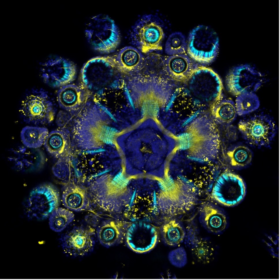
Laurent Formery’s image of a sea urchin won an honorable mention in our Image of the Year Award 2020.
Q: What do you find most fascinating about microscopy?
Laurent: Microscopy is a window to a different world. It reveals the beauty of small things that we overlook by looking with our bare eyes. What I find the most fascinating is to take a sample of sea water (with a plankton net, ideally) and look at it under a compound microscope. It’s teeming with life. Some of these microscopic algae, larvae, and animals that you see have incredible shapes, colors, or forms. A fascinating world that you would have no idea about without microscopes. Microscopes also show things that are more abstract. In the case of fluorescence imaging, microscopes reveal structures that have a beauty of their own, such as a nervous system.
Q: Where does this fascination stem from?
Laurent: I have always found that plants, flowers, and insects are beautiful and fascinating. Now that I’m a scientist and have access to microscopes, I can also appreciate the beauty of life at a much smaller scale—plankton, for instance, or small invertebrates.
Laurent Formery’s image of a sea urchin won an honorable mention in our Image of the Year Award 2020.
Q: Does your profession intersect with imaging? Or is this more of a hobby, art form, or passion for you?
Laurent: I am now a postdoctoral researcher in the Lowe Laboratory at Stanford University. I study the evolution of animals from a molecular perspective, trying to understand how modifications of ancient genetic systems enabled the evolution of animal groups with completely different anatomies. Imaging is key to my research. I usually spend around 10 hours a week using microscopes (confocal microscopes for the most part).
Q: What are you currently working on professionally?
Laurent: My research is about the evolution of body plans in animals. I am focusing on invertebrate groups that are closely related to us, in particular echinoderms (sea stars, sea urchins, sea cucumbers). Although they look very different to us, these animals form their adult body using the same genetic tools as us (they use the same architect genes).
Differences in their adult anatomy comes from differences in the wiring of these same genes during their development. Recently, we have been working on how the genes that create the antero-posterior axis in vertebrates have been modified in echinoderms to accommodate their unique pentaradial symmetry. This work is now available as a preprint and will be published soon.
To keep it short, vertebrates pattern their antero-posterior axis to make distinct regions of the body, such as the head and the trunk. In echinoderms, we found that only head genes are activated, while a trunk is completely missing. In a way, echinoderms like sea stars should be seen as walking heads creeping on the sea floor.
Be the Star of Our Next Image of the Year Award
Stay tuned for our Image of the Year contest announcement next month! Our light microscopy imaging competition continues to expand, and we can’t wait to see how you capture the art of science through the microscope lens.
Related Content
Image of the Year 2022 Americas Winner Captures the Art of the Natural World
Small Masterpieces of Nature—Meet the Image of the Year 2022 EMEA Winner
Image of the Year 2022 APAC Winner Captures the Colorful Microscopic World