
The new year has kicked off, and we’re celebrating by sharing a series of new (and extinct) samples captured under the microscope. From diatoms to dinosaurs, here are the most-liked microscope images for January 2022.
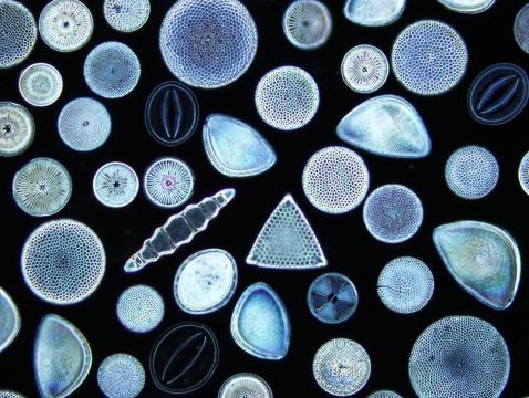
Our top image for the month is a Victorian-arranged diatom slide dating back to the 1880s that was imaged using modern Olympus technology. Dr. Hunter Hines used darkfield microscopy and magnified the image 200x to show off the shells of these single cells.
Image courtesy of Hunter Hines. 2020 Image of the Year Award submission. Captured using an Olympus BX53 microscope with a DP72 camera.
Our 2021 Image of the Year competition has been extended and is accepting entries through February 28! Learn more and submit your images at olympus-lifescience.com/ioty.
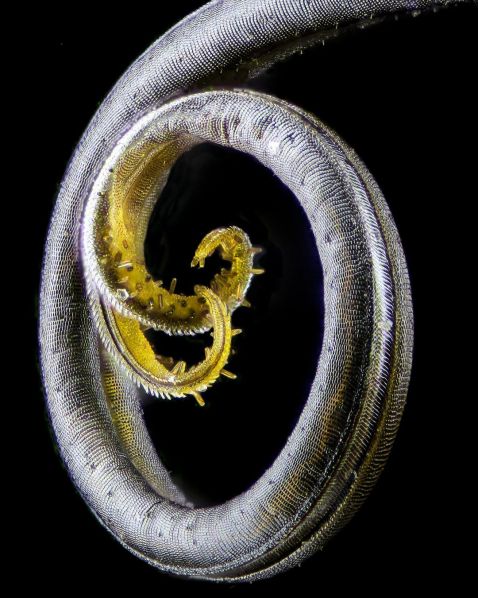
This fern-like curlicue is actually a close-up view of the proboscis of a Virginia ctenucha moth. A moth's proboscis is normally tightly coiled (as pictured here), but they uncoil to their full length to suck nectar or other fluids.
Image courtesy of Caleb Foster. 2020 Image of the Year Award submission. Captured using an Olympus 4X PLAPO objective; Z-projection of a 180-image Z-stack.
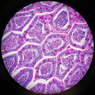
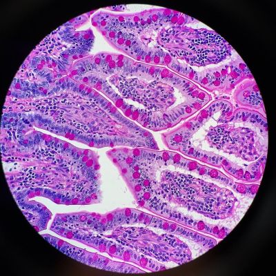
As part of her “Special Stain Saturday” series, Kate Murphy imaged a section of a dog’s small intestine.
Kate explains, “Today’s stain is Periodic Acid–Schiff (PAS), and it stains mucin a bright magenta color. Mucins are glycoproteins that provide lubrication within our gastrointestinal tract. PAS will stain for all mucins. And it can be paired with alcian blue to help differentiate acid from neutral mucin. Neutral mucins are found in the stomach. Acid mucins are found in the small intestine.”
Image and caption courtesy of Kate Murphy. Captured using an Olympus BX40 microscope.
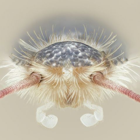
Captured by Marco Jongsma, this popular image takes a close look at a silverfish.
“The silverfish or sugar guest (Lepisma saccharina) is a small insect belonging to the order Zygentoma. Its name refers to the silvery sheen of the scales that cover the body and perhaps also to its ability to slip at lightning speed. Silverfish are about one centimeter long and, in the Netherlands and Belgium, are mainly found in damp corners in bathrooms or kitchen cupboards,” Marco explains.
The insect seems much cuter up close than when they crawl across our sinks, don’t you agree?
Image and caption courtesy of Marco Jongsma. Captured using an Olympus BH2 microscope.
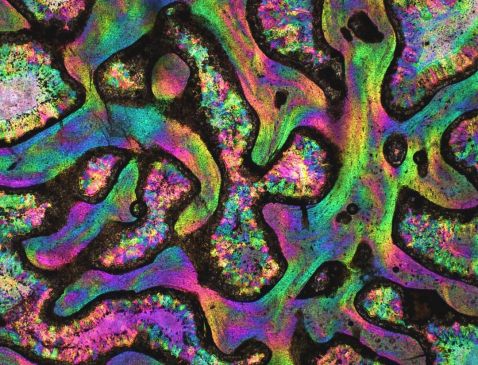
Ever wonder what dinosaur bone looks like under the microscope? This image is a magnified look at dinosaur bone with polychromatic polarization add-on. It was captured using illuminating white polarized light with a spectral fan of polarization ellipses. The colors captured in this vibrant image are the same colors seen by the naked eye.
Image courtesy of Michael Shribak. 2020 Image of the Year Award submission. Captured using an Olympus IX83 microscope with a 4X objective.
Bonus video! We’re a little bit jelly of how mesmerizing this video is.
“This is a jellyfish baby, the so-called Ephyra. It’s a developmental stage in the jellyfish lifecycle that comes right before the mature medusa. The medusa larva settles down on a substrate, developing into a polyp. The polyp will grow into a strobila, which is the budding stage releasing the above-mentioned Ephyra into the water. The Ephyra will then develop into the mature jellyfish medusa.”
Video courtesy of Benedikt Pleyer. Captured on an Olympus BX53 microscope at 400x magnification with 1000x brightfield.
To see more images like these, be sure to follow us on Instagram at @olympuslifescience.
Interested in sharing your own images? Visit our image submission site or enter our global imaging contest!
Related Content
Surf and Turf—Our Most Popular Microscope Images for December 2021
Aesthetics of Autofluorescence—Meet the IOTY 2020 Global Winner
Lifelong Love of the Microscopic—Meet the IOTY 2020 Americas Regional Winner
